 Movie
Movie Controller

[English] 日本語
 Yorodumi
- EMDB-2962: Cryo-electron microscopy structure of the unoccupied population o... -
+ Open data

- Basic information
| Entry | Database: EMDB / ID: EMD-2962 | |||||||||
|---|---|---|---|---|---|---|---|---|---|---|
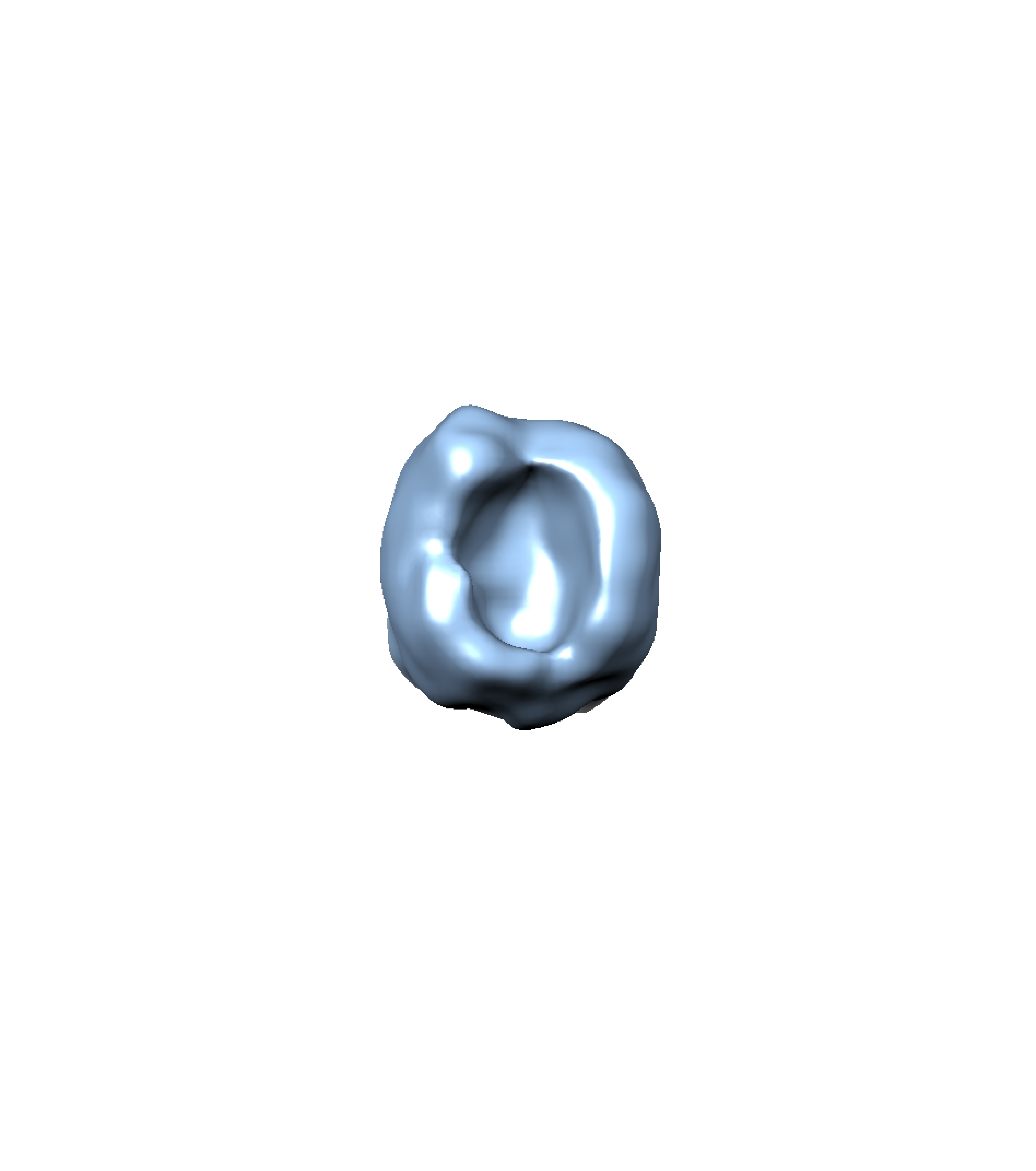
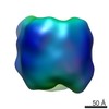
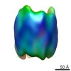
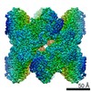
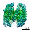
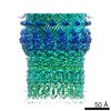
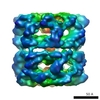
| Title | Cryo-electron microscopy structure of the unoccupied population of CCT5 complexes after incubation with mHtt | |||||||||
 Map data Map data | Single particle tomography reconstruction of unoccupied CCT5 complex after incubation with mutant huntington. | |||||||||
 Sample Sample |
| |||||||||
 Keywords Keywords | chaperonin / Huntington's disease / protein aggregation | |||||||||
| Function / homology |  Function and homology information Function and homology informationpositive regulation of establishment of protein localization to telomere / positive regulation of protein localization to Cajal body / BBSome-mediated cargo-targeting to cilium / binding of sperm to zona pellucida / Folding of actin by CCT/TriC / Formation of tubulin folding intermediates by CCT/TriC / positive regulation of telomerase RNA localization to Cajal body / chaperonin-containing T-complex / Prefoldin mediated transfer of substrate to CCT/TriC / beta-tubulin binding ...positive regulation of establishment of protein localization to telomere / positive regulation of protein localization to Cajal body / BBSome-mediated cargo-targeting to cilium / binding of sperm to zona pellucida / Folding of actin by CCT/TriC / Formation of tubulin folding intermediates by CCT/TriC / positive regulation of telomerase RNA localization to Cajal body / chaperonin-containing T-complex / Prefoldin mediated transfer of substrate to CCT/TriC / beta-tubulin binding / Association of TriC/CCT with target proteins during biosynthesis / chaperone-mediated protein folding / protein folding chaperone / positive regulation of telomere maintenance via telomerase / mRNA 3'-UTR binding / ATP-dependent protein folding chaperone / response to virus / mRNA 5'-UTR binding / Cooperation of PDCL (PhLP1) and TRiC/CCT in G-protein beta folding / G-protein beta-subunit binding / unfolded protein binding / protein folding / cell body / microtubule / protein stabilization / centrosome / ATP hydrolysis activity / extracellular exosome / ATP binding / cytosol Similarity search - Function | |||||||||
| Biological species |  Homo sapiens (human) Homo sapiens (human) | |||||||||
| Method | subtomogram averaging / cryo EM | |||||||||
 Authors Authors | Darrow MC / Sergeeva OA / Isas JM / Galaz-Montoya J / King JA / Langen R / Schmid MF / Chiu W | |||||||||
 Citation Citation | Journal: J Biol Chem / Year: 2014 Title: Biochemical characterization of mutants in chaperonin proteins CCT4 and CCT5 associated with hereditary sensory neuropathy. Authors: Oksana A Sergeeva / Meme T Tran / Cameron Haase-Pettingell / Jonathan A King /  Abstract: Hereditary sensory neuropathies are a class of disorders marked by degeneration of the nerve fibers in the sensory periphery neurons. Recently, two mutations were identified in the subunits of the ...Hereditary sensory neuropathies are a class of disorders marked by degeneration of the nerve fibers in the sensory periphery neurons. Recently, two mutations were identified in the subunits of the eukaryotic cytosolic chaperonin TRiC, a protein machine responsible for folding actin and tubulin in the cell. C450Y CCT4 was identified in a stock of Sprague-Dawley rats, whereas H147R CCT5 was found in a human Moroccan family. As with many genetically identified mutations associated with neuropathies, the underlying molecular basis of the mutants was not defined. We investigated the biochemical properties of these mutants using an expression system in Escherichia coli that produces homo-oligomeric rings of CCT4 and CCT5. Full-length versions of both mutant protein chains were expressed in E. coli at levels approaching that of the WT chains. Sucrose gradient centrifugation revealed chaperonin-sized complexes of both WT and mutant chaperonins, but with reduced recovery of C450Y CCT4 soluble subunits. Electron microscopy of negatively stained samples of C450Y CCT4 revealed few ring-shaped species, whereas WT CCT4, H147R CCT5, and WT CCT5 revealed similar ring structures. CCT5 complexes were assayed for their ability to suppress aggregation of and refold the model substrate γd-crystallin, suppress aggregation of mutant huntingtin, and refold the physiological substrate β-actin in vitro. H147R CCT5 was not as efficient in chaperoning these substrates as WT CCT5. The subtle effects of these mutations are consistent with the homozygous disease phenotype, in which most functions are carried out during development and adulthood, but some selective function is lost or reduced. | |||||||||
| History |
|
- Structure visualization
| Movie |
Movie viewer |
|---|---|
| Structure viewer | EM map: SurfViewMolmilJmol/JSmol |
| Supplemental images |
- Downloads & links
-EMDB archive
| Map data | emd_2962.map.gz | 1.3 MB | EMDB map data format | |
|---|---|---|---|---|
| Header (meta data) | emd-2962-v30.xmlemd-2962.xml | 14 KB 14 KB | Display Display | EMDB header |
| Images | emd_2962.tif | 128.1 MB | ||
| Archive directory |  http://ftp.pdbj.org/pub/emdb/structures/EMD-2962ftp://ftp.pdbj.org/pub/emdb/structures/EMD-2962 http://ftp.pdbj.org/pub/emdb/structures/EMD-2962ftp://ftp.pdbj.org/pub/emdb/structures/EMD-2962 | HTTPS FTP |
-Validation report
| Summary document | emd_2962_validation.pdf.gz | 205.5 KB | Display | EMDB validaton report |
|---|---|---|---|---|
| Full document | emd_2962_full_validation.pdf.gz | 204.6 KB | Display | |
| Data in XML | emd_2962_validation.xml.gz | 5.2 KB | Display | |
| Arichive directory | https://ftp.pdbj.org/pub/emdb/validation_reports/EMD-2962ftp://ftp.pdbj.org/pub/emdb/validation_reports/EMD-2962 | HTTPS FTP |
-Related structure data
-Links
| EMDB pages | EMDB (EBI/PDBe) / EMDataResource |
|---|---|
| Related items in Molecule of the Month |


-Map
| File | Download / File: emd_2962.map.gz / Format: CCP4 / Size: 1.4 MB / Type: IMAGE STORED AS FLOATING POINT NUMBER (4 BYTES) | ||||||||||||||||||||||||||||||||||||||||||||||||||||||||||||||||||||
|---|---|---|---|---|---|---|---|---|---|---|---|---|---|---|---|---|---|---|---|---|---|---|---|---|---|---|---|---|---|---|---|---|---|---|---|---|---|---|---|---|---|---|---|---|---|---|---|---|---|---|---|---|---|---|---|---|---|---|---|---|---|---|---|---|---|---|---|---|---|
| Annotation | Single particle tomography reconstruction of unoccupied CCT5 complex after incubation with mutant huntington. | ||||||||||||||||||||||||||||||||||||||||||||||||||||||||||||||||||||
| Voxel size | X=Y=Z: 4.704 Å | ||||||||||||||||||||||||||||||||||||||||||||||||||||||||||||||||||||
| Density |
| ||||||||||||||||||||||||||||||||||||||||||||||||||||||||||||||||||||
| Symmetry | Space group: 1 | ||||||||||||||||||||||||||||||||||||||||||||||||||||||||||||||||||||
| Details | EMDB XML:
CCP4 map header:
| ||||||||||||||||||||||||||||||||||||||||||||||||||||||||||||||||||||
-Supplemental data
- Sample components
-Entire : Unoccupied CCT5 complex after incubation with mutant huntingtin e...
| Entire | Name: Unoccupied CCT5 complex after incubation with mutant huntingtin exon 1. |
|---|---|
| Components |
|
-Supramolecule #1000: Unoccupied CCT5 complex after incubation with mutant huntingtin e...
| Supramolecule | Name: Unoccupied CCT5 complex after incubation with mutant huntingtin exon 1. type: sample / ID: 1000 Oligomeric state: 16 CCT5 subunits make up one CCT5 complex. Number unique components: 1 |
|---|
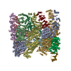
-Macromolecule #1: chaperonin containing TCP1, subunit 5 (epsilon) complex
| Macromolecule | Name: chaperonin containing TCP1, subunit 5 (epsilon) complex type: protein_or_peptide / ID: 1 / Name.synonym: CCT5 Complex / Oligomeric state: Hexadecamer / Recombinant expression: Yes |
|---|---|
| Source (natural) | Organism: Homo sapiens (human) / synonym: Human / Location in cell: cytoplasm |
| Molecular weight | Experimental: 1.0 MDa / Theoretical: 960 KDa |
| Recombinant expression | Organism:  |
| Sequence | UniProtKB: T-complex protein 1 subunit epsilon / GO: chaperonin-containing T-complex, protein folding / InterPro: Chaperonin Cpn60/GroEL/TCP-1 family |
-Experimental details
-Structure determination
| Method | cryo EM |
|---|---|
 Processing Processing | subtomogram averaging |
| Aggregation state | particle |
-Sample preparation
| Concentration | 0.8 mg/mL |
|---|---|
| Buffer | pH: 8 / Details: 20 mM Tris, 150 mM NaCl, 1 mM DTT, 1% glycerol |
| Grid | Details: 200 mesh Quantifoil copper grid R 2/2 holey carbon support, glow discharged |
| Vitrification | Cryogen name: ETHANE / Chamber humidity: 85 % / Chamber temperature: 95 K / Instrument: FEI VITROBOT MARK III Method: 1x or 2x double sided blotting for 1 or 2 seconds each blot before plunging. |
- Electron microscopy #1
| Microscopy ID | 1 |
|---|---|
| Microscope | JEOL 2200FS |
| Temperature | Average: 95 K |
| Specialist optics | Energy filter - Name: Gatan / Energy filter - Lower energy threshold: 0.0 eV / Energy filter - Upper energy threshold: 25.0 eV |
| Date | Feb 12, 2014 |
| Image recording | Category: CCD / Film or detector model: GATAN ULTRASCAN 4000 (4k x 4k) / Average electron dose: 2.0 e/Å2 / Camera length: 1200 |
| Electron beam | Acceleration voltage: 200 kV / Electron source:  FIELD EMISSION GUN FIELD EMISSION GUN |
| Electron optics | Illumination mode: FLOOD BEAM / Imaging mode: BRIGHT FIELD / Cs: 2.0 mm / Nominal defocus max: 5.0 µm / Nominal defocus min: 5.0 µm / Nominal magnification: 25000 |
| Sample stage | Specimen holder: LN2 cooled / Specimen holder model: GATAN LIQUID NITROGEN / Tilt series - Axis1 - Min angle: -55 ° / Tilt series - Axis1 - Max angle: 55 ° |
-Electron microscopy #2
| Microscopy ID | 2 |
|---|---|
| Microscope | JEOL 2200FS |
| Temperature | Average: 95 K |
| Specialist optics | Energy filter - Name: Gatan / Energy filter - Lower energy threshold: 0.0 eV / Energy filter - Upper energy threshold: 25.0 eV |
| Date | Oct 31, 2013 |
| Image recording | Category: CCD / Film or detector model: GATAN ULTRASCAN 4000 (4k x 4k) / Average electron dose: 2.0 e/Å2 / Camera length: 1200 |
| Electron beam | Acceleration voltage: 200 kV / Electron source: FIELD EMISSION GUN |
| Electron optics | Illumination mode: FLOOD BEAM / Imaging mode: BRIGHT FIELD / Cs: 2.0 mm / Nominal defocus max: 5.0 µm / Nominal defocus min: 5.0 µm / Nominal magnification: 25000 |
| Sample stage | Specimen holder: LN2 cooled / Specimen holder model: GATAN LIQUID NITROGEN / Tilt series - Axis1 - Min angle: -55 ° / Tilt series - Axis1 - Max angle: 55 ° |
-Image processing
| Details | Subvolumes were manually selected using IMOD for coordinates and EMAN2 for boxing. Further processing was completed in EMAN2. |
|---|---|
| Final reconstruction | Applied symmetry - Point group: C1 (asymmetric) / Algorithm: OTHER / Software - Name: IMOD, EMAN2 / Number subtomograms used: 300 |
