 Movie
Movie Controller


+ Open data

- Basic information
| Entry | Database: EMDB / ID: EMD-2599 | |||||||||
|---|---|---|---|---|---|---|---|---|---|---|
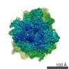
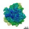
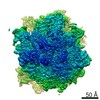
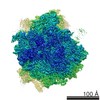
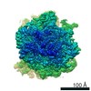
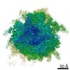
| Title | Kluyveromyces lactis 80S ribosome in complex with CrPV-IRES | |||||||||
 Map data Map data | Kluyveromyces lactis 80S ribosome in complex with CrPV-IRES in canonical state | |||||||||
 Sample Sample |
| |||||||||
 Keywords Keywords |  eukaryotic / translation / initiation / ribosome / IRES eukaryotic / translation / initiation / ribosome / IRES | |||||||||
| Biological species |  Kluyveromyces lactis (yeast) / Kluyveromyces lactis (yeast) /  Cricket paralysis virus Cricket paralysis virus | |||||||||
| Method | single particle reconstruction / cryo EM / Resolution: 3.7 Å | |||||||||
 Authors Authors | Fernandez IS / Bai XC / Scheres SHW / Ramakrishnan V | |||||||||
 Citation Citation | Journal: Cell / Year: 2014 Title: Initiation of translation by cricket paralysis virus IRES requires its translocation in the ribosome. Authors: Israel S Fernández / Xiao-Chen Bai / Garib Murshudov / Sjors H W Scheres / V Ramakrishnan /  Abstract: The cricket paralysis virus internal ribosome entry site (CrPV-IRES) is a folded structure in a viral mRNA that allows initiation of translation in the absence of any host initiation factors. By ...The cricket paralysis virus internal ribosome entry site (CrPV-IRES) is a folded structure in a viral mRNA that allows initiation of translation in the absence of any host initiation factors. By using recent advances in single-particle electron cryomicroscopy, we have solved the structure of CrPV-IRES bound to the ribosome of the yeast Kluyveromyces lactis in both the canonical and rotated states at overall resolutions of 3.7 and 3.8 Å, respectively. In both states, the pseudoknot PKI of the CrPV-IRES mimics a tRNA/mRNA interaction in the decoding center of the A site of the 40S ribosomal subunit. The structure and accompanying factor-binding data show that CrPV-IRES binding mimics a pretranslocation rather than initiation state of the ribosome. Translocation of the IRES by elongation factor 2 (eEF2) is required to bring the first codon of the mRNA into the A site and to allow the start of translation. | |||||||||
| History |
|
- Structure visualization
| Movie |
 Movie viewer Movie viewer |
|---|---|
| Structure viewer | EM map: SurfViewMolmilJmol/JSmol |
| Supplemental images |

- Downloads & links
-EMDB archive
| Map data | emd_2599.map.gz | 20.8 MB | EMDB map data format | |
|---|---|---|---|---|
| Header (meta data) | emd-2599-v30.xmlemd-2599.xml | 19.3 KB 19.3 KB | Display Display | EMDB header |
| Images | 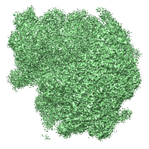 image2599.png image2599.png | 389.9 KB | ||
| Others | 40S_IntRat_WM.map80S_IRES_IntRat_FINAL.map | 125 MB 125 MB | ||
| Archive directory |  http://ftp.pdbj.org/pub/emdb/structures/EMD-2599ftp://ftp.pdbj.org/pub/emdb/structures/EMD-2599 http://ftp.pdbj.org/pub/emdb/structures/EMD-2599ftp://ftp.pdbj.org/pub/emdb/structures/EMD-2599 | HTTPS FTP |
-Related structure data
| Related structure data |  4v91MC  2603C  2604C  4v92C M: atomic model generated by this map C: citing same article ( |
|---|---|
| Similar structure data |


-Links
| EMDB pages | EMDB (EBI/PDBe) / EMDataResource |
|---|---|
| Related items in Molecule of the Month |

-Map
| File | Download / File: emd_2599.map.gz / Format: CCP4 / Size: 122.1 MB / Type: IMAGE STORED AS FLOATING POINT NUMBER (4 BYTES) | ||||||||||||||||||||||||||||||||||||||||||||||||||||||||||||
|---|---|---|---|---|---|---|---|---|---|---|---|---|---|---|---|---|---|---|---|---|---|---|---|---|---|---|---|---|---|---|---|---|---|---|---|---|---|---|---|---|---|---|---|---|---|---|---|---|---|---|---|---|---|---|---|---|---|---|---|---|---|
| Annotation | Kluyveromyces lactis 80S ribosome in complex with CrPV-IRES in canonical state | ||||||||||||||||||||||||||||||||||||||||||||||||||||||||||||
| Voxel size | X=Y=Z: 1.34 Å | ||||||||||||||||||||||||||||||||||||||||||||||||||||||||||||
| Density |
| ||||||||||||||||||||||||||||||||||||||||||||||||||||||||||||
| Symmetry | Space group: 1 | ||||||||||||||||||||||||||||||||||||||||||||||||||||||||||||
| Details | EMDB XML:
CCP4 map header:
| ||||||||||||||||||||||||||||||||||||||||||||||||||||||||||||
-Supplemental data
-Supplemental map: 40S IntRat WM.map
| File | 40S_IntRat_WM.map | ||||||||||||
|---|---|---|---|---|---|---|---|---|---|---|---|---|---|
| Projections & Slices |
| ||||||||||||
| Density Histograms |
 Z
Z Y
Y X
X


-Supplemental map: 80S IRES IntRat FINAL.map
| File | 80S_IRES_IntRat_FINAL.map | ||||||||||||
|---|---|---|---|---|---|---|---|---|---|---|---|---|---|
| Projections & Slices |
| ||||||||||||
| Density Histograms |


- Sample components
-Entire : Kluyveromyces lactis 80S ribosome in complex with CrPV-IRES
| Entire | Name: Kluyveromyces lactis 80S ribosome in complex with CrPV-IRES |
|---|---|
| Components |
|
-Supramolecule #1000: Kluyveromyces lactis 80S ribosome in complex with CrPV-IRES
| Supramolecule | Name: Kluyveromyces lactis 80S ribosome in complex with CrPV-IRES type: sample / ID: 1000 / Details: The sample was monodisperse / Oligomeric state: 1 / Number unique components: 2 |
|---|---|
| Molecular weight | Experimental: 3.2 MDa / Theoretical: 3.2 MDa / Method: Sedimentation |
-Supramolecule #1: Kluyveromyces lactis 80S ribosome
| Supramolecule | Name: Kluyveromyces lactis 80S ribosome / type: complex / ID: 1 / Name.synonym: Kluyveromyces lactis 80S ribosome / Recombinant expression: No / Ribosome-details: ribosome-eukaryote: ALL |
|---|---|
| Source (natural) | Organism: Kluyveromyces lactis (yeast) |
| Molecular weight | Experimental: 3.2 MDa / Theoretical: 3.2 MDa |
-Macromolecule #1: IRES
| Macromolecule | Name: IRES / type: protein_or_peptide / ID: 1 / Recombinant expression: No |
|---|---|
| Source (natural) | Organism: Cricket paralysis virus |
-Experimental details
-Structure determination
| Method | cryo EM |
|---|---|
 Processing Processing | single particle reconstruction |
| Aggregation state | particle |
-Sample preparation
| Concentration | 0.1 mg/mL |
|---|---|
| Buffer | pH: 6.5 Details: 10mM MES-KOH, 10mM NH4 acetate, 40mM K-acetate, 8 mM Mg-acetated, 2mM DTT |
| Grid | Details: 400 mesh R 2/2 Quantifoil grids |
| Vitrification | Cryogen name: PROPANE / Chamber humidity: 90 % / Chamber temperature: 120 K / Instrument: FEI VITROBOT MARK I Timed resolved state: Vitrified 30 msec after spraying with effector Method: 2.5 |
- Electron microscopy
| Microscope | FEI TITAN KRIOS |
|---|---|
| Electron beam | Acceleration voltage: 300 kV / Electron source: FIELD EMISSION GUN |
| Electron optics | Illumination mode: OTHER / Imaging mode: BRIGHT FIELDBright-field microscopy / Cs: 2.7 mm / Nominal defocus max: 0.003 µm / Nominal defocus min: 0.0018 µm / Nominal magnification: 47000 |
| Sample stage | Specimen holder model: FEI TITAN KRIOS AUTOGRID HOLDER |
| Date | Jul 7, 2013 |
| Image recording | Category: CCD / Film or detector model: FEI FALCON II (4k x 4k) / Number real images: 1900 / Average electron dose: 40 e/Å2 |
| Experimental equipment |  Model: Titan Krios / Image courtesy: FEI Company |
-Image processing
| CTF correction | Details: Each particle |
|---|---|
| Final reconstruction | Applied symmetry - Point group: C1 (asymmetric) / Algorithm: OTHER / Resolution.type: BY AUTHOR / Resolution: 3.7 Å / Resolution method: OTHER / Software - Name: Relion / Number images used: 18132 |
| Details | RELION movement correction processing used |
-Atomic model buiding 1
| Initial model | PDB ID: Chain - #0 - Chain ID: A / Chain - #1 - Chain ID: B |
|---|---|
| Software | Name: Chimera, refmac |
| Details | Coordinates refined with Refmac and checked with COOT |
| Refinement | Space: RECIPROCAL / Protocol: FLEXIBLE FIT / Overall B value: 60 / Target criteria: R-factor, FSC |
| Output model | PDB-4v91: |

-Atomic model buiding 2
| Initial model | PDB ID: Chain - Chain ID: A |
|---|---|
| Software | Name: Chimera, refmac |
| Details | Coordinates refined with Refmac and checked with COOT |
| Refinement | Space: RECIPROCAL / Protocol: FLEXIBLE FIT / Overall B value: 60 / Target criteria: R-factor, FSC |
| Output model | PDB-4v91: |
-Atomic model buiding 3
| Initial model | PDB ID:  3u5c Chain - #0 - Chain ID: A / Chain - #1 - Chain ID: B / Chain - #2 - Chain ID: C / Chain - #3 - Chain ID: D / Chain - #4 - Chain ID: E / Chain - #5 - Chain ID: F / Chain - #6 - Chain ID: G / Chain - #7 - Chain ID: H / Chain - #8 - Chain ID: I / Chain - #9 - Chain ID: J / Chain - #10 - Chain ID: K / Chain - #11 - Chain ID: L / Chain - #12 - Chain ID: M / Chain - #13 - Chain ID: N / Chain - #14 - Chain ID: O / Chain - #15 - Chain ID: P / Chain - #16 - Chain ID: Q / Chain - #17 - Chain ID: R / Chain - #18 - Chain ID: S / Chain - #19 - Chain ID: T / Chain - #20 - Chain ID: U / Chain - #21 - Chain ID: V / Chain - #22 - Chain ID: W / Chain - #23 - Chain ID: X / Chain - #24 - Chain ID: Y / Chain - #25 - Chain ID: Z / Chain - #26 - Chain ID: a / Chain - #27 - Chain ID: b / Chain - #28 - Chain ID: c / Chain - #29 - Chain ID: d / Chain - #30 - Chain ID: e / Chain - #31 - Chain ID: f / Chain - #32 - Chain ID: g |
|---|---|
| Software | Name: Chimera, refmac |
| Details | Coordinates refined with Refmac and checked with COOT |
| Refinement | Space: RECIPROCAL / Protocol: FLEXIBLE FIT / Overall B value: 60 / Target criteria: R-factor, FSC |
| Output model | PDB-4v91: |
-Atomic model buiding 4
| Initial model | PDB ID: 3u5e Chain - #0 - Chain ID: A / Chain - #1 - Chain ID: B / Chain - #2 - Chain ID: C / Chain - #3 - Chain ID: D / Chain - #4 - Chain ID: E / Chain - #5 - Chain ID: F / Chain - #6 - Chain ID: G / Chain - #7 - Chain ID: H / Chain - #8 - Chain ID: I / Chain - #9 - Chain ID: J / Chain - #10 - Chain ID: K / Chain - #11 - Chain ID: L / Chain - #12 - Chain ID: M / Chain - #13 - Chain ID: N / Chain - #14 - Chain ID: O / Chain - #15 - Chain ID: P / Chain - #16 - Chain ID: Q / Chain - #17 - Chain ID: R / Chain - #18 - Chain ID: S / Chain - #19 - Chain ID: T / Chain - #20 - Chain ID: U / Chain - #21 - Chain ID: V / Chain - #22 - Chain ID: W / Chain - #23 - Chain ID: X / Chain - #24 - Chain ID: Y / Chain - #25 - Chain ID: Z / Chain - #26 - Chain ID: a / Chain - #27 - Chain ID: b / Chain - #28 - Chain ID: c / Chain - #29 - Chain ID: d / Chain - #30 - Chain ID: e / Chain - #31 - Chain ID: f / Chain - #32 - Chain ID: g / Chain - #33 - Chain ID: h / Chain - #34 - Chain ID: i / Chain - #35 - Chain ID: j / Chain - #36 - Chain ID: k / Chain - #37 - Chain ID: l / Chain - #38 - Chain ID: m / Chain - #39 - Chain ID: n / Chain - #40 - Chain ID: o / Chain - #41 - Chain ID: p |
|---|---|
| Software | Name: Chimera, refmac |
| Details | Coordinates refined with Refmac and checked with COOT |
| Refinement | Space: RECIPROCAL / Protocol: FLEXIBLE FIT / Overall B value: 60 / Target criteria: R-factor, FSC |
| Output model | PDB-4v91: |
-Atomic model buiding 5
| Initial model | PDB ID: 3u5d Chain - #0 - Chain ID: 1 / Chain - #1 - Chain ID: 3 / Chain - #2 - Chain ID: 4 |
|---|---|
| Software | Name: Chimera, refmac |
| Details | Coordinates refined with Refmac and checked with COOT |
| Refinement | Space: RECIPROCAL / Protocol: FLEXIBLE FIT / Overall B value: 60 / Target criteria: R-factor, FSC |
| Output model | PDB-4v91: |
