 Movie
Movie Controller

[English] 日本語
 Yorodumi
- EMDB-1957: Cryo Electron Tomography of Herpes Simplex Virus during Axonal Tr... -
+ Open data

- Basic information
| Entry | Database: EMDB / ID: EMD-1957 | |||||||||
|---|---|---|---|---|---|---|---|---|---|---|
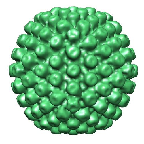
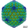
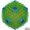
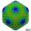
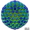
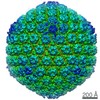
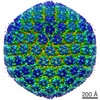
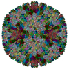
| Title | Cryo Electron Tomography of Herpes Simplex Virus during Axonal Transport and Secondary Envelopment in Primary Neurons | |||||||||
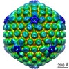
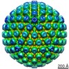
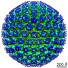
 Map data Map data | Cytosolic AB capsid | |||||||||
 Sample Sample |
| |||||||||
 Keywords Keywords | HSV-1 / HSV1 / herpesvirus / herpes simplex virus 1 / capsid / cytosolic A/B-capsid / tegument | |||||||||
| Biological species |   Human herpesvirus 1 (Herpes simplex virus type 1) Human herpesvirus 1 (Herpes simplex virus type 1) | |||||||||
| Method | subtomogram averaging / cryo EM / negative staining / Resolution: 97.0 Å | |||||||||
 Authors Authors | Ibiricu I / Huiskonen JT / Doehner K / Bradke F / Sodeik B / Gruenewald K | |||||||||
 Citation Citation | Journal: PLoS Pathog / Year: 2011 Title: Cryo electron tomography of herpes simplex virus during axonal transport and secondary envelopment in primary neurons. Authors: Iosune Ibiricu / Juha T Huiskonen / Katinka Döhner / Frank Bradke / Beate Sodeik / Kay Grünewald /  Abstract: During herpes simplex virus 1 (HSV1) egress in neurons, viral particles travel from the neuronal cell body along the axon towards the synapse. Whether HSV1 particles are transported as enveloped ...During herpes simplex virus 1 (HSV1) egress in neurons, viral particles travel from the neuronal cell body along the axon towards the synapse. Whether HSV1 particles are transported as enveloped virions as proposed by the 'married' model or as non-enveloped capsids suggested by the 'separate' model is controversial. Specific viral proteins may form a recruitment platform for microtubule motors that catalyze such transport. However, their subviral location has remained elusive. Here we established a system to analyze herpesvirus egress by cryo electron tomography. At 16 h post infection, we observed intra-axonal transport of progeny HSV1 viral particles in dissociated hippocampal neurons by live-cell fluorescence microscopy. Cryo electron tomography of frozen-hydrated neurons revealed that most egressing capsids were transported independently of the viral envelope. Unexpectedly, we found not only DNA-containing capsids (cytosolic C-capsids), but also capsids lacking DNA (cytosolic A-/B-capsids) in mid-axon regions. Subvolume averaging revealed lower amounts of tegument on cytosolic A-/B-capsids than on C-capsids. Nevertheless, all capsid types underwent active axonal transport. Therefore, even few tegument proteins on the capsid vertices seemed to suffice for transport. Secondary envelopment of capsids was observed at axon terminals. On their luminal face, the enveloping vesicles were studded with typical glycoprotein-like spikes. Furthermore, we noted an accretion of tegument density at the concave cytosolic face of the vesicle membrane in close proximity to the capsids. Three-dimensional analysis revealed that these assembly sites lacked cytoskeletal elements, but that filamentous actin surrounded them and formed an assembly compartment. Our data support the 'separate model' for HSV1 egress, i.e. progeny herpes viruses being transported along axons as subassemblies and not as complete virions within transport vesicles. | |||||||||
| History |
|
- Structure visualization
| Movie |
 Movie viewer Movie viewer |
|---|---|
| Structure viewer | EM map: SurfViewMolmilJmol/JSmol |
| Supplemental images |
- Downloads & links
-EMDB archive
| Map data | emd_1957.map.gz | 24.9 MB | EMDB map data format | |
|---|---|---|---|---|
| Header (meta data) | emd-1957-v30.xmlemd-1957.xml | 9.8 KB 9.8 KB | Display Display | EMDB header |
| Images | EMD_1957_cytosolic_ab.tif | 298.7 KB | ||
| Archive directory |  http://ftp.pdbj.org/pub/emdb/structures/EMD-1957ftp://ftp.pdbj.org/pub/emdb/structures/EMD-1957 http://ftp.pdbj.org/pub/emdb/structures/EMD-1957ftp://ftp.pdbj.org/pub/emdb/structures/EMD-1957 | HTTPS FTP |
-Validation report
| Summary document | emd_1957_validation.pdf.gz | 258.5 KB | Display | EMDB validaton report |
|---|---|---|---|---|
| Full document | emd_1957_full_validation.pdf.gz | 257.7 KB | Display | |
| Data in XML | emd_1957_validation.xml.gz | 6.2 KB | Display | |
| Arichive directory | https://ftp.pdbj.org/pub/emdb/validation_reports/EMD-1957ftp://ftp.pdbj.org/pub/emdb/validation_reports/EMD-1957 | HTTPS FTP |
-Related structure data

-Links
| EMDB pages | EMDB (EBI/PDBe) / EMDataResource |
|---|
-Map
| File | Download / File: emd_1957.map.gz / Format: CCP4 / Size: 29.8 MB / Type: IMAGE STORED AS FLOATING POINT NUMBER (4 BYTES) | ||||||||||||||||||||||||||||||||||||||||||||||||||||||||||||||||||||
|---|---|---|---|---|---|---|---|---|---|---|---|---|---|---|---|---|---|---|---|---|---|---|---|---|---|---|---|---|---|---|---|---|---|---|---|---|---|---|---|---|---|---|---|---|---|---|---|---|---|---|---|---|---|---|---|---|---|---|---|---|---|---|---|---|---|---|---|---|---|
| Annotation | Cytosolic AB capsid | ||||||||||||||||||||||||||||||||||||||||||||||||||||||||||||||||||||
| Voxel size | X=Y=Z: 8.05 Å | ||||||||||||||||||||||||||||||||||||||||||||||||||||||||||||||||||||
| Density |
| ||||||||||||||||||||||||||||||||||||||||||||||||||||||||||||||||||||
| Symmetry | Space group: 1 | ||||||||||||||||||||||||||||||||||||||||||||||||||||||||||||||||||||
| Details | EMDB XML:
CCP4 map header:
| ||||||||||||||||||||||||||||||||||||||||||||||||||||||||||||||||||||
-Supplemental data
- Sample components
-Entire : Herpes simplex virus 1 cytosolic AB-capsid
| Entire | Name: Herpes simplex virus 1 cytosolic AB-capsid |
|---|---|
| Components |
|
-Supramolecule #1000: Herpes simplex virus 1 cytosolic AB-capsid
| Supramolecule | Name: Herpes simplex virus 1 cytosolic AB-capsid / type: sample / ID: 1000 / Oligomeric state: Icosahedral Capsid / Number unique components: 1 |
|---|
-Supramolecule #1: Human herpesvirus 1
| Supramolecule | Name: Human herpesvirus 1 / type: virus / ID: 1 / Name.synonym: Herpes simplex virus 1 / NCBI-ID: 10298 / Sci species name: Human herpesvirus 1 / Database: NCBI / Virus type: VIRUS-LIKE PARTICLE / Virus isolate: STRAIN / Virus enveloped: No / Virus empty: No / Syn species name: Herpes simplex virus 1 |
|---|---|
| Host (natural) | Organism:  |
| Virus shell | Shell ID: 1 / Name: capsid / Diameter: 1250 Å / T number (triangulation number): 16 |
-Experimental details
-Structure determination
| Method | negative staining, cryo EM |
|---|---|
 Processing Processing | subtomogram averaging |
| Aggregation state | particle |
-Sample preparation
| Buffer | pH: 7.4 |
|---|---|
| Staining | Type: NEGATIVE / Details: Unstained |
| Grid | Details: 200 mesh Au grids with holey carbon support film (Au R2/1 200 mesh, Quantifoil |
| Vitrification | Cryogen name: ETHANE / Instrument: OTHER / Method: Manual blotting |
- Electron microscopy
| Microscope | FEI POLARA 300 |
|---|---|
| Specialist optics | Energy filter - Name: GIF2002 / Energy filter - Lower energy threshold: 0.0 eV / Energy filter - Upper energy threshold: 10.0 eV |
| Details | Tilt series |
| Image recording | Category: CCD / Film or detector model: GENERIC CCD / Average electron dose: 80 e/Å2 |
| Electron beam | Acceleration voltage: 300 kV / Electron source:  FIELD EMISSION GUN FIELD EMISSION GUN |
| Electron optics | Calibrated magnification: 37267 / Illumination mode: FLOOD BEAM / Imaging mode: BRIGHT FIELD / Cs: 2.0 mm / Nominal defocus max: 12.0 µm / Nominal defocus min: 10.0 µm / Nominal magnification: 27500 |
| Sample stage | Specimen holder: Eucentric / Specimen holder model: OTHER / Tilt series - Axis1 - Min angle: -60 ° / Tilt series - Axis1 - Max angle: 60 ° |
| Experimental equipment |  Model: Tecnai Polara / Image courtesy: FEI Company |
-Image processing
| Details | Sub-volumes of icosahedral particles where manually picked in tomographic reconstructions, aligned and averaged. Average number of projections used in the 3D reconstructions: 26. |
|---|---|
| Final reconstruction | Algorithm: OTHER / Resolution.type: BY AUTHOR / Resolution: 97.0 Å / Resolution method: FSC 0.5 CUT-OFF / Software - Name: IMOD and Bsoft / Details: Map was calculated from tomographic sub-volumes |
| CTF correction | Details: Low pass filter |
