Movie
Movie Controller


+ Open data

- Basic information
| Entry | Database: EMDB / ID: EMD-1735 | |||||||||
|---|---|---|---|---|---|---|---|---|---|---|
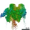
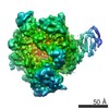
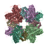
| Title | Rubisco in complex with Rubisco large subunit methyltransferase | |||||||||
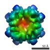
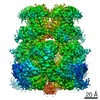
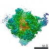
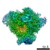
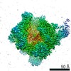
 Map data Map data | This is a volume of RuBisCO in complex with RuBisCO large subunit methyl transferase | |||||||||
 Sample Sample |
| |||||||||
 Keywords Keywords | Rubisco / carbon fixation / methylation | |||||||||
| Biological species |  Spinacia oleracea (spinach) / Spinacia oleracea (spinach) /  Pisum sativum (garden pea) Pisum sativum (garden pea) | |||||||||
| Method | single particle reconstruction / cryo EM / Resolution: 11.0 Å | |||||||||
 Authors Authors | Raunser S / Magnani R / Huang Z / Houtz RL / Trievel RC / Penczek PA / Walz T | |||||||||
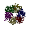
 Citation Citation | Journal: Proc Natl Acad Sci U S A / Year: 2009 Title: Rubisco in complex with Rubisco large subunit methyltransferase. Authors: Stefan Raunser / Roberta Magnani / Zhong Huang / Robert L Houtz / Raymond C Trievel / Pawel A Penczek / Thomas Walz /  Abstract: SET domain protein lysine methyltransferases (PKMT) are a structurally unique class of enzymes that catalyze the specific methylation of lysine residues in a number of different substrates. ...SET domain protein lysine methyltransferases (PKMT) are a structurally unique class of enzymes that catalyze the specific methylation of lysine residues in a number of different substrates. Especially histone-specific SET domain PKMTs have received widespread attention because of their roles in the regulation of epigenetic gene expression and the development of some cancers. Rubisco large subunit methyltransferase (RLSMT) is a chloroplast-localized SET domain PKMT responsible for the formation of trimethyl-lysine-14 in the large subunit of Rubisco, an essential photosynthetic enzyme. Here, we have used cryoelectron microscopy to produce an 11-A density map of the Rubisco-RLSMT complex. The atomic model of the complex, obtained by fitting crystal structures of Rubisco and RLSMT into the density map, shows that the extensive contact regions between the 2 proteins are mainly mediated by hydrophobic residues and leucine-rich repeats. It further provides insights into potential conformational changes that may occur during substrate binding and catalysis. This study presents the first structural analysis of a SET domain PKMT in complex with its intact polypeptide substrate. | |||||||||
| History |
|
- Structure visualization
| Movie |
 Movie viewer Movie viewer |
|---|---|
| Structure viewer | EM map: SurfViewMolmilJmol/JSmol |
| Supplemental images |

- Downloads & links
-EMDB archive
| Map data | emd_1735.map.gz | 1.9 MB | EMDB map data format | |
|---|---|---|---|---|
| Header (meta data) | emd-1735-v30.xmlemd-1735.xml | 11.7 KB 11.7 KB | Display Display | EMDB header |
| Images | EMD-1735.tif | 763.8 KB | ||
| Archive directory |  http://ftp.pdbj.org/pub/emdb/structures/EMD-1735ftp://ftp.pdbj.org/pub/emdb/structures/EMD-1735 http://ftp.pdbj.org/pub/emdb/structures/EMD-1735ftp://ftp.pdbj.org/pub/emdb/structures/EMD-1735 | HTTPS FTP |
-Related structure data
-Links
| EMDB pages | EMDB (EBI/PDBe) / EMDataResource |
|---|
-Map
| File | Download / File: emd_1735.map.gz / Format: CCP4 / Size: 1.9 MB / Type: IMAGE STORED AS FLOATING POINT NUMBER (4 BYTES) | ||||||||||||||||||||||||||||||||||||||||||||||||||||||||||||||||||||
|---|---|---|---|---|---|---|---|---|---|---|---|---|---|---|---|---|---|---|---|---|---|---|---|---|---|---|---|---|---|---|---|---|---|---|---|---|---|---|---|---|---|---|---|---|---|---|---|---|---|---|---|---|---|---|---|---|---|---|---|---|---|---|---|---|---|---|---|---|---|
| Annotation | This is a volume of RuBisCO in complex with RuBisCO large subunit methyl transferase | ||||||||||||||||||||||||||||||||||||||||||||||||||||||||||||||||||||
| Voxel size | X=Y=Z: 4.1 Å | ||||||||||||||||||||||||||||||||||||||||||||||||||||||||||||||||||||
| Density |
| ||||||||||||||||||||||||||||||||||||||||||||||||||||||||||||||||||||
| Symmetry | Space group: 1 | ||||||||||||||||||||||||||||||||||||||||||||||||||||||||||||||||||||
| Details | EMDB XML:
CCP4 map header:
| ||||||||||||||||||||||||||||||||||||||||||||||||||||||||||||||||||||
-Supplemental data
- Sample components
-Entire : Spinach RuBisCO in complex with pea RuBisCO LSMT
| Entire | Name: Spinach RuBisCO in complex with pea RuBisCO LSMT |
|---|---|
| Components |
|
-Supramolecule #1000: Spinach RuBisCO in complex with pea RuBisCO LSMT
| Supramolecule | Name: Spinach RuBisCO in complex with pea RuBisCO LSMT / type: sample / ID: 1000 Details: The sample was thawed from storage at -80 degrees Celcius before being loaded onto the grid. Oligomeric state: One monomer of LSMT binds to a RuBisCO hexadecamer Number unique components: 2 |
|---|---|
| Molecular weight | Experimental: 585 KDa / Theoretical: 585 KDa |
-Macromolecule #1: Ribulose-1,5-bisphosphate carboxylase oxygenase
| Macromolecule | Name: Ribulose-1,5-bisphosphate carboxylase oxygenase / type: protein_or_peptide / ID: 1 / Name.synonym: RuBisCO / Number of copies: 1 / Oligomeric state: Hexadecamer / Recombinant expression: No |
|---|---|
| Source (natural) | Organism: Spinacia oleracea (spinach) / synonym: Spinach / Tissue: Plant leaves / Organelle: Chloroplast / Location in cell: Stroma |
| Molecular weight | Experimental: 534 KDa / Theoretical: 534 KDa |
-Macromolecule #2: RuBisCO large subunit methyl transferase
| Macromolecule | Name: RuBisCO large subunit methyl transferase / type: protein_or_peptide / ID: 2 / Name.synonym: RLSMT / Number of copies: 1 / Oligomeric state: Monomer / Recombinant expression: Yes |
|---|---|
| Source (natural) | Organism: Pisum sativum (garden pea) / synonym: Pea / Tissue: Plant leaves / Organelle: Chloroplast / Location in cell: Stroma |
| Molecular weight | Experimental: 55 KDa / Theoretical: 55 KDa |
| Recombinant expression | Organism:  Escherichia coli (E. coli) / Recombinant plasmid: pDEST14 Escherichia coli (E. coli) / Recombinant plasmid: pDEST14 |
-Experimental details
-Structure determination
| Method | cryo EM |
|---|---|
 Processing Processing | single particle reconstruction |
| Aggregation state | particle |
-Sample preparation
| Concentration | 5 mg/mL |
|---|---|
| Buffer | pH: 8 / Details: 50 mM Tris-HCl pH 8, 5 mM MgCl2, 1 mM EDTA |
| Grid | Details: Quantifoil grids 2 um holes |
| Vitrification | Cryogen name: ETHANE / Chamber humidity: 90 % / Instrument: OTHER / Details: Vitrification instrument: Vitrobot / Method: Blot for 1 s before plunging |
- Electron microscopy
| Microscope | FEI TECNAI F20 |
|---|---|
| Electron beam | Acceleration voltage: 200 kV / Electron source: FIELD EMISSION GUN |
| Electron optics | Calibrated magnification: 51159 / Illumination mode: FLOOD BEAM / Imaging mode: BRIGHT FIELDBright-field microscopy / Cs: 2.0 mm / Nominal defocus max: 5.0 µm / Nominal defocus min: 2.5 µm / Nominal magnification: 50000 |
| Sample stage | Specimen holder: Side entry liquid nitrogen-cooled cryo specimen holder. Specimen holder model: OTHER |
| Image recording | Category: FILM / Film or detector model: KODAK SO-163 FILM / Digitization - Scanner: ZEISS SCAI / Digitization - Sampling interval: 7 µm / Number real images: 110 / Average electron dose: 15 e/Å2 |
| Tilt angle min | 0 |
| Tilt angle max | 0 |
| Experimental equipment |  Model: Tecnai F20 / Image courtesy: FEI Company |
-Image processing
| CTF correction | Details: Each particle |
|---|---|
| Final angle assignment | Details: SPIDER:theta 45 degrees, phi 45 degrees |
| Final reconstruction | Applied symmetry - Point group: C1 (asymmetric) / Algorithm: OTHER / Resolution.type: BY AUTHOR / Resolution: 11.0 Å / Resolution method: FSC 0.5 CUT-OFF / Software - Name: SPARX / Number images used: 29000 |
| Details | The particles were selected interactively at the computer terminal. |
-Atomic model buiding 1
| Initial model | PDB ID: |
|---|---|
| Software | Name: SITUS |
| Details | Protocol: Rigid Body |
| Refinement | Space: REAL / Protocol: RIGID BODY FIT / Target criteria: R-factor |
-Atomic model buiding 2
| Initial model | PDB ID: |
|---|---|
| Software | Name: SITUS |
| Details | Protocol: Rigid Body |
| Refinement | Space: REAL / Protocol: RIGID BODY FIT / Target criteria: R-factor |