 Movie
Movie Controller

[English] 日本語
 Yorodumi
- EMDB-1688: CryoEM reconstruction of human parechovirus 1 complexed with inte... -
+ Open data

- Basic information
| Entry | Database: EMDB / ID: EMD-1688 | |||||||||
|---|---|---|---|---|---|---|---|---|---|---|
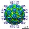
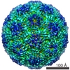
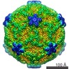
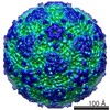
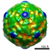
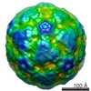
| Title | CryoEM reconstruction of human parechovirus 1 complexed with integrin alphaV-beta3 | |||||||||
 Map data Map data | This is a cryoEM reconstruction of human parechovirus 1 complexed with integrin alphaV-beta3 at 15 angstrom resolution | |||||||||
 Sample Sample |
| |||||||||
 Keywords Keywords |  picornavirus / parechovirus / icosahedral / integrin picornavirus / parechovirus / icosahedral / integrin | |||||||||
| Biological species |  Homo sapiens (human) / Homo sapiens (human) /  Human parechovirus 1 Human parechovirus 1 | |||||||||
| Method | single particle reconstruction / cryo EM / negative staining / Resolution: 15.0 Å | |||||||||
 Authors Authors | Seitsonen JJT / Susi P / Heikkila O / Laurinmaki P / Hyypia T / Butcher SJ | |||||||||
 Citation Citation | Journal: J Virol / Year: 2010 Title: Interaction of alphaVbeta3 and alphaVbeta6 integrins with human parechovirus 1. Authors: Jani Seitsonen / Petri Susi / Outi Heikkilä / Robert S Sinkovits / Pasi Laurinmäki / Timo Hyypiä / Sarah J Butcher /  Abstract: Human parechovirus (HPEV) infections are very common in early childhood and can be severe in neonates. It has been shown that integrins are important for cellular infectivity of HPEV1 through ...Human parechovirus (HPEV) infections are very common in early childhood and can be severe in neonates. It has been shown that integrins are important for cellular infectivity of HPEV1 through experiments using peptide blocking assays and function-blocking antibodies to alpha(V) integrins. The interaction of HPEV1 with alpha(V) integrins is presumably mediated by a C-terminal RGD motif in the capsid protein VP1. We characterized the binding of integrins alpha(V)beta(3) and alpha(V)beta(6) to HPEV1 by biochemical and structural studies. We showed that although HPEV1 bound efficiently to immobilized integrins, alpha(V)beta(6) bound more efficiently than alpha(V)beta(3) to immobilized HPEV1. Moreover, soluble alpha(V)beta(6), but not alpha(V)beta(3), blocked HPEV1 cellular infectivity, indicating that it is a high-affinity receptor for HPEV1. We also showed that HPEV1 binding to integrins in vitro could be partially blocked by RGD peptides. Using electron cryo-microscopy and image reconstruction, we showed that HPEV1 has the typical T=1 (pseudo T=3) organization of a picornavirus. Complexes of HPEV1 and integrins indicated that both integrin footprints reside between the 5-fold and 3-fold symmetry axes. This result does not match the RGD position predicted from the coxsackievirus A9 X-ray structure but is consistent with the predicted location of this motif in the shorter C terminus found in HPEV1. This first structural characterization of a parechovirus indicates that the differences in receptor binding are due to the amino acid differences in the integrins rather than to significantly different viral footprints. | |||||||||
| History |
|
- Structure visualization
| Movie |
 Movie viewer Movie viewer |
|---|---|
| Structure viewer | EM map: SurfViewMolmilJmol/JSmol |
| Supplemental images |

- Downloads & links
-EMDB archive
| Map data | emd_1688.map.gz | 40.3 MB | EMDB map data format | |
|---|---|---|---|---|
| Header (meta data) | emd-1688-v30.xmlemd-1688.xml | 10.4 KB 10.4 KB | Display Display | EMDB header |
| Images | EMD-1688_HPEV1avb3.tif | 732.6 KB | ||
| Archive directory |  http://ftp.pdbj.org/pub/emdb/structures/EMD-1688ftp://ftp.pdbj.org/pub/emdb/structures/EMD-1688 http://ftp.pdbj.org/pub/emdb/structures/EMD-1688ftp://ftp.pdbj.org/pub/emdb/structures/EMD-1688 | HTTPS FTP |
-Related structure data


-Links
| EMDB pages | EMDB (EBI/PDBe) / EMDataResource |
|---|
-Map
| File | Download / File: emd_1688.map.gz / Format: CCP4 / Size: 120.1 MB / Type: IMAGE STORED AS SIGNED INTEGER (2 BYTES) | ||||||||||||||||||||||||||||||||||||||||||||||||||||||||||||||||||||
|---|---|---|---|---|---|---|---|---|---|---|---|---|---|---|---|---|---|---|---|---|---|---|---|---|---|---|---|---|---|---|---|---|---|---|---|---|---|---|---|---|---|---|---|---|---|---|---|---|---|---|---|---|---|---|---|---|---|---|---|---|---|---|---|---|---|---|---|---|---|
| Annotation | This is a cryoEM reconstruction of human parechovirus 1 complexed with integrin alphaV-beta3 at 15 angstrom resolution | ||||||||||||||||||||||||||||||||||||||||||||||||||||||||||||||||||||
| Projections & slices | Image control
Images are generated by Spider. | ||||||||||||||||||||||||||||||||||||||||||||||||||||||||||||||||||||
| Voxel size | X=Y=Z: 1.13 Å | ||||||||||||||||||||||||||||||||||||||||||||||||||||||||||||||||||||
| Density |
| ||||||||||||||||||||||||||||||||||||||||||||||||||||||||||||||||||||
| Symmetry | Space group: 1 | ||||||||||||||||||||||||||||||||||||||||||||||||||||||||||||||||||||
| Details | EMDB XML:
CCP4 map header:
| ||||||||||||||||||||||||||||||||||||||||||||||||||||||||||||||||||||
 Z (Sec.)
 Y (Row.)
 X (Col.)


-Supplemental data
- Sample components
-Entire : Purified human parechovirus 1 in 1 mM MgCl2 and integrin alphaV-b...
| Entire | Name: Purified human parechovirus 1 in 1 mM MgCl2 and integrin alphaV-beta6 in PBS buffer |
|---|---|
| Components |
|
-Supramolecule #1000: Purified human parechovirus 1 in 1 mM MgCl2 and integrin alphaV-b...
| Supramolecule | Name: Purified human parechovirus 1 in 1 mM MgCl2 and integrin alphaV-beta6 in PBS buffer type: sample / ID: 1000 Oligomeric state: Complete particle (60-mer) with the RNA genome inside with few integrin molecules attached Number unique components: 2 |
|---|
-Supramolecule #1: Human parechovirus 1
| Supramolecule | Name: Human parechovirus 1 / type: virus / ID: 1 / Name.synonym: HPEV1 / NCBI-ID: 12063 / Sci species name: Human parechovirus 1 / Virus type: VIRION / Virus isolate: STRAIN / Virus enveloped: No / Virus empty: No / Syn species name: HPEV1 |
|---|---|
| Host (natural) | Organism: Homo sapiens (human) / synonym: VERTEBRATES |
| Molecular weight | Theoretical: 5.1 MDa |
| Virus shell | Shell ID: 1 / Name: native / Diameter: 280 Å / T number (triangulation number): 1 |
-Macromolecule #1: Integrin alphaV-beta3
| Macromolecule | Name: Integrin alphaV-beta3 / type: protein_or_peptide / ID: 1 / Name.synonym: Integrin alphaV-beta3 / Details: Product of BioMarket Ltd., Finland / Number of copies: 1 / Oligomeric state: Heterodimer / Recombinant expression: Yes |
|---|---|
| Source (natural) | Organism: Homo sapiens (human) / synonym: Human / Location in cell: Plasma membrane |
| Molecular weight | Theoretical: 203 KDa |
-Experimental details
-Structure determination
| Method | negative staining, cryo EM |
|---|---|
 Processing Processing | single particle reconstruction |
| Aggregation state | particle |
-Sample preparation
| Concentration | 1.24 mg/mL |
|---|---|
| Buffer | pH: 7.2 / Details: 1 mM MgCl2 in PBS |
| Staining | Type: NEGATIVE / Details: Cryo preparation |
| Grid | Details: 400 mesh carbon coated copper |
| Vitrification | Cryogen name: ETHANE / Instrument: HOMEMADE PLUNGER / Details: Vitrification instrument: Guillotine / Method: Blot for 1-2 seconds before plunging |
- Electron microscopy
| Microscope | FEI TECNAI F20 |
|---|---|
| Electron beam | Acceleration voltage: 200 kV / Electron source: FIELD EMISSION GUN |
| Electron optics | Calibrated magnification: 60500 / Illumination mode: FLOOD BEAM / Imaging mode: BRIGHT FIELDBright-field microscopy / Cs: 2.0 mm / Nominal defocus max: 2.922 µm / Nominal defocus min: 1.486 µm / Nominal magnification: 62000 |
| Sample stage | Specimen holder: Gatan 626 / Specimen holder model: GATAN LIQUID NITROGEN |
| Temperature | Average: 91 K |
| Alignment procedure | Legacy - Astigmatism: objective lens astigmatism was corrected at 62,000 times magnification |
| Image recording | Category: FILM / Film or detector model: KODAK SO-163 FILM / Digitization - Scanner: OTHER / Digitization - Sampling interval: 7 µm / Number real images: 16 / Average electron dose: 18 e/Å2 / Details: Scanned with Zeiss Photoscan TD scanner |
| Experimental equipment |  Model: Tecnai F20 / Image courtesy: FEI Company |
-Image processing
| CTF correction | Details: Each micrograph |
|---|---|
| Final reconstruction | Applied symmetry - Point group: I (icosahedral) / Algorithm: OTHER / Resolution.type: BY AUTHOR / Resolution: 15.0 Å / Resolution method: FSC 0.5 CUT-OFF / Software - Name: AUTO3DEM / Number images used: 431 |
