 Movie
Movie Controller

[English] 日本語
 Yorodumi
- EMDB-1648: Assembly and Allosteric Mechanism of Molluscan Hemocyanin Reveale... -
+ Open data

- Basic information
| Entry | Database: EMDB / ID: EMD-1648 | |||||||||
|---|---|---|---|---|---|---|---|---|---|---|
| Title | Assembly and Allosteric Mechanism of Molluscan Hemocyanin Revealed by Cryo-EM Structure and Pseudo-atomic Model | |||||||||
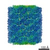
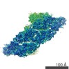
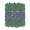
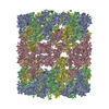
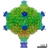
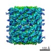
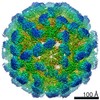
 Map data Map data | The whole structure of Haliotis diversicolor Hemocyanin isoform 1 (HdH1) is a hollow cylindrical dodecamer. Each of its 20 subunits is composed of 8 functional units (FUs). | |||||||||
 Sample Sample |
| |||||||||
 Keywords Keywords |  Oxygen binding / allosteric mechanism Oxygen binding / allosteric mechanism | |||||||||
| Function / homology | Di-copper centre-containing domain superfamily / metabolic process Function and homology information Function and homology information | |||||||||
| Biological species |  Haliotis diversicolor (invertebrata) Haliotis diversicolor (invertebrata) | |||||||||
| Method | single particle reconstruction / cryo EM / Resolution: 7.8 Å | |||||||||
 Authors Authors | Xinghong D / Junjie Z / Jiangyong W / Kunpeng L / Donghua C / Qinfen Z / Wah C | |||||||||
 Citation Citation | Journal: Structure / Year: 2013 Title: Cryo-EM structure of a molluscan hemocyanin suggests its allosteric mechanism. Authors: Qinfen Zhang / Xinghong Dai / Yao Cong / Junjie Zhang / Dong-Hua Chen / Matthew T Dougherty / Jiangyong Wang / Steven J Ludtke / Michael F Schmid / Wah Chiu /  Abstract: Hemocyanins are responsible for transporting O2 in the arthropod and molluscan hemolymph. Haliotis diversicolor molluscan hemocyanin isoform 1 (HdH1) is an 8 MDa oligomer. Each subunit is made up of ...Hemocyanins are responsible for transporting O2 in the arthropod and molluscan hemolymph. Haliotis diversicolor molluscan hemocyanin isoform 1 (HdH1) is an 8 MDa oligomer. Each subunit is made up of eight functional units (FUs). Each FU contains two Cu ions, which can reversibly bind an oxygen molecule. Here, we report a 4.5 A° cryo-EM structure of HdH1. The structure clearly shows ten asymmetric units arranged with D5 symmetry. Each asymmetric unit contains two structurally distinct but chemically identical subunits. The map is sufficiently resolved to trace the entire subunit Ca backbone and to visualize densities corresponding to some large side chains, Cu ion pairs, and interaction networks of adjacent subunits. A FU topology path intertwining between the two subunits of the asymmetric unit is unambiguously determined. Our observations suggest a structural mechanism for the stability of the entire hemocyanin didecamer and 20 ‘‘communication clusters’’ across asymmetric units responsible for its allosteric property upon oxygen binding. | |||||||||
| History |
|
- Structure visualization
| Movie |
Movie viewer |
|---|---|
| Structure viewer | EM map: SurfViewMolmilJmol/JSmol |
| Supplemental images |

- Downloads & links
-EMDB archive
| Map data | emd_1648.map.gz | 277.7 MB | EMDB map data format | |
|---|---|---|---|---|
| Header (meta data) | emd-1648-v30.xmlemd-1648.xml | 10.1 KB 10.1 KB | Display Display | EMDB header |
| Images | 1648_A-1.tif | 762.3 KB | ||
| Archive directory |  http://ftp.pdbj.org/pub/emdb/structures/EMD-1648ftp://ftp.pdbj.org/pub/emdb/structures/EMD-1648 http://ftp.pdbj.org/pub/emdb/structures/EMD-1648ftp://ftp.pdbj.org/pub/emdb/structures/EMD-1648 | HTTPS FTP |
-Related structure data
-Links
| EMDB pages | EMDB (EBI/PDBe) / EMDataResource |
|---|
-Map
| File | Download / File: emd_1648.map.gz / Format: CCP4 / Size: 300.3 MB / Type: IMAGE STORED AS FLOATING POINT NUMBER (4 BYTES) | ||||||||||||||||||||||||||||||||||||||||||||||||||||||||||||
|---|---|---|---|---|---|---|---|---|---|---|---|---|---|---|---|---|---|---|---|---|---|---|---|---|---|---|---|---|---|---|---|---|---|---|---|---|---|---|---|---|---|---|---|---|---|---|---|---|---|---|---|---|---|---|---|---|---|---|---|---|---|
| Annotation | The whole structure of Haliotis diversicolor Hemocyanin isoform 1 (HdH1) is a hollow cylindrical dodecamer. Each of its 20 subunits is composed of 8 functional units (FUs). | ||||||||||||||||||||||||||||||||||||||||||||||||||||||||||||
| Projections & slices | Image control
Images are generated by Spider. | ||||||||||||||||||||||||||||||||||||||||||||||||||||||||||||
| Voxel size | X=Y=Z: 1.06 Å | ||||||||||||||||||||||||||||||||||||||||||||||||||||||||||||
| Density |
| ||||||||||||||||||||||||||||||||||||||||||||||||||||||||||||
| Symmetry | Space group: 1 | ||||||||||||||||||||||||||||||||||||||||||||||||||||||||||||
| Details | EMDB XML:
CCP4 map header:
| ||||||||||||||||||||||||||||||||||||||||||||||||||||||||||||
 Z (Sec.)
 Y (Row.)
 X (Col.)


-Supplemental data
- Sample components
-Entire : Haliotis diversicolor (Gastropod, Mollusca) Hemocyanin isoform 1 ...
| Entire | Name: Haliotis diversicolor (Gastropod, Mollusca) Hemocyanin isoform 1 (HdH1) |
|---|---|
| Components |
|
-Supramolecule #1000: Haliotis diversicolor (Gastropod, Mollusca) Hemocyanin isoform 1 ...
| Supramolecule | Name: Haliotis diversicolor (Gastropod, Mollusca) Hemocyanin isoform 1 (HdH1) type: sample / ID: 1000 / Oligomeric state: Dodecamer / Number unique components: 1 |
|---|---|
| Molecular weight | Experimental: 8 MDa / Theoretical: 8 MDa / Method: SDS-PAGE |
-Macromolecule #1: Hemocyanin
| Macromolecule | Name: Hemocyanin / type: protein_or_peptide / ID: 1 / Name.synonym: Hemocyanin / Number of copies: 20 / Oligomeric state: Dodecamer / Recombinant expression: No |
|---|---|
| Source (natural) | Organism: Haliotis diversicolor (invertebrata) / Tissue: Blood |
| Molecular weight | Experimental: 8 MDa / Theoretical: 8 MDa |
| Sequence | GO: metabolic process / InterPro: Di-copper centre-containing domain superfamily |
-Experimental details
-Structure determination
| Method | cryo EM |
|---|---|
 Processing Processing | single particle reconstruction |
| Aggregation state | particle |
-Sample preparation
| Buffer | pH: 7.5 Details: 0.2M NaCl , 50mM Tris-HCl, 5mM CaCl2, 5mM MgCl2, pH7.5 |
|---|---|
| Grid | Details: 1.2/1.3 copper Quantifoil grids |
| Vitrification | Cryogen name: ETHANE / Chamber humidity: 100 % / Chamber temperature: 101 K / Instrument: OTHER / Details: Vitrification instrument: Vitrobot / Method: Blot for 2 seconds before plunging |
- Electron microscopy
| Microscope | JEOL 3200FSC |
|---|---|
| Electron beam | Acceleration voltage: 300 kV / Electron source: FIELD EMISSION GUN |
| Electron optics | Illumination mode: FLOOD BEAM / Imaging mode: BRIGHT FIELDBright-field microscopy / Cs: 4.1 mm / Nominal defocus max: 3.5 µm / Nominal defocus min: 0.8 µm / Nominal magnification: 60000 |
| Specialist optics | Energy filter - Name: JEOL in-column |
| Sample stage | Specimen holder: Eucentric / Specimen holder model: JEOL 3200FSC CRYOHOLDER |
| Temperature | Average: 101 K |
| Alignment procedure | Legacy - Astigmatism: Objective lens astigmatism was corrected at 400,000 times magnification |
| Details | MDS |
| Image recording | Category: FILM / Film or detector model: KODAK SO-163 FILM / Digitization - Scanner: NIKON SUPER COOLSCAN 9000 / Digitization - Sampling interval: 6.35 µm / Number real images: 820 / Average electron dose: 18 e/Å2 / Bits/pixel: 16 |
-Image processing
| CTF correction | Details: Each micrograph |
|---|---|
| Final reconstruction | Applied symmetry - Point group: D5 (2x5 fold dihedral) / Resolution.type: BY AUTHOR / Resolution: 7.8 Å / Resolution method: FSC 0.5 CUT-OFF / Software - Name: EMAN / Number images used: 41650 |
| Details | Particles were automatically boxed out from micrographs by e2boxer.py from EMAN2 single particle analysis software package, and CTF correction was carried out with EMAN program CTFIT. All the 3D reconstruction was done with EMAN1.8. |
