 Movie
Movie Controller

[English] 日本語
 Yorodumi
- EMDB-1347: The eukaryotic translation initiation factors eIF1 and eIF1A indu... -
+ Open data

- Basic information
| Entry | Database: EMDB / ID: EMD-1347 | |||||||||
|---|---|---|---|---|---|---|---|---|---|---|
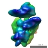
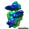
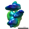
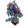
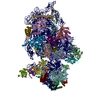
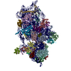
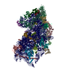
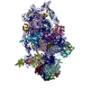
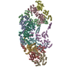
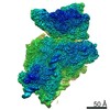
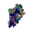
| Title | The eukaryotic translation initiation factors eIF1 and eIF1A induce an open conformation of the 40S ribosome. | |||||||||
 Map data Map data | Yeast 40S small ribosomal subunit bound to the translation initiation factors eIF1 and eIF1A | |||||||||
 Sample Sample |
| |||||||||
| Function / homology |  SUI1 domain / Translation initiation factor 1A (eIF-1A) / translation initiation factor activity SUI1 domain / Translation initiation factor 1A (eIF-1A) / translation initiation factor activity Function and homology information Function and homology information | |||||||||
| Biological species |  Saccharomyces cerevisiae (brewer's yeast) Saccharomyces cerevisiae (brewer's yeast) | |||||||||
| Method | single particle reconstruction / cryo EM / Resolution: 22.0 Å | |||||||||
 Authors Authors | Passmore LA / Schmeing TM / Maag D / Applefield DJ / Acker MG / Algire MA / Lorsch JR / Ramakrishnan V | |||||||||
 Citation Citation | Journal: Mol Cell / Year: 2007 Title: The eukaryotic translation initiation factors eIF1 and eIF1A induce an open conformation of the 40S ribosome. Authors: Lori A Passmore / T Martin Schmeing / David Maag / Drew J Applefield / Michael G Acker / Mikkel A Algire / Jon R Lorsch / V Ramakrishnan /  Abstract: Initiation of translation is the process by which initiator tRNA and the start codon of mRNA are positioned in the ribosomal P site. In eukaryotes, one of the first steps involves the binding of two ...Initiation of translation is the process by which initiator tRNA and the start codon of mRNA are positioned in the ribosomal P site. In eukaryotes, one of the first steps involves the binding of two small factors, eIF1 and eIF1A, to the small (40S) ribosomal subunit. This facilitates tRNA binding, allows scanning of mRNA, and maintains fidelity of start codon recognition. Using cryo-EM, we have obtained 3D reconstructions of 40S bound to both eIF1 and eIF1A, and with each factor alone. These structures reveal that together, eIF1 and eIF1A stabilize a conformational change that opens the mRNA binding channel. Biochemical data reveal that both factors accelerate the rate of ternary complex (eIF2*GTP*Met-tRNA(i)(Met)) binding to 40S but only eIF1A stabilizes this interaction. Our results suggest that eIF1 and eIF1A promote an open, scanning-competent preinitiation complex that closes upon start codon recognition and eIF1 release to stabilize ternary complex binding and clamp down on mRNA. | |||||||||
| History |
|
- Structure visualization
| Movie |
Movie viewer |
|---|---|
| Structure viewer | EM map: SurfViewMolmilJmol/JSmol |
| Supplemental images |

- Downloads & links
-EMDB archive
| Map data | emd_1347.map.gz | 14 MB | EMDB map data format | |
|---|---|---|---|---|
| Header (meta data) | emd-1347-v30.xmlemd-1347.xml | 12.5 KB 12.5 KB | Display Display | EMDB header |
| Images |  1347.gif 1347.gif | 86.4 KB | ||
| Archive directory |  http://ftp.pdbj.org/pub/emdb/structures/EMD-1347ftp://ftp.pdbj.org/pub/emdb/structures/EMD-1347 http://ftp.pdbj.org/pub/emdb/structures/EMD-1347ftp://ftp.pdbj.org/pub/emdb/structures/EMD-1347 | HTTPS FTP |
-Related structure data
-Links
| EMDB pages | EMDB (EBI/PDBe) / EMDataResource |
|---|---|
| Related items in Molecule of the Month |

-Map
| File | Download / File: emd_1347.map.gz / Format: CCP4 / Size: 26.4 MB / Type: IMAGE STORED AS FLOATING POINT NUMBER (4 BYTES) | ||||||||||||||||||||||||||||||||||||||||||||||||||||||||||||||||||||
|---|---|---|---|---|---|---|---|---|---|---|---|---|---|---|---|---|---|---|---|---|---|---|---|---|---|---|---|---|---|---|---|---|---|---|---|---|---|---|---|---|---|---|---|---|---|---|---|---|---|---|---|---|---|---|---|---|---|---|---|---|---|---|---|---|---|---|---|---|---|
| Annotation | Yeast 40S small ribosomal subunit bound to the translation initiation factors eIF1 and eIF1A | ||||||||||||||||||||||||||||||||||||||||||||||||||||||||||||||||||||
| Projections & slices | Image control
Images are generated by Spider. | ||||||||||||||||||||||||||||||||||||||||||||||||||||||||||||||||||||
| Voxel size | X=Y=Z: 2.4 Å | ||||||||||||||||||||||||||||||||||||||||||||||||||||||||||||||||||||
| Density |
| ||||||||||||||||||||||||||||||||||||||||||||||||||||||||||||||||||||
| Symmetry | Space group: 1 | ||||||||||||||||||||||||||||||||||||||||||||||||||||||||||||||||||||
| Details | EMDB XML:
CCP4 map header:
| ||||||||||||||||||||||||||||||||||||||||||||||||||||||||||||||||||||
 Z (Sec.)
 Y (Row.)
 X (Col.)


-Supplemental data
- Sample components
-Entire : S. cerevisiae 40S ribosomal subunit bound to eIF1 and eIF1A
| Entire | Name: S. cerevisiae 40S ribosomal subunit bound to eIF1 and eIF1A |
|---|---|
| Components |
|
-Supramolecule #1000: S. cerevisiae 40S ribosomal subunit bound to eIF1 and eIF1A
| Supramolecule | Name: S. cerevisiae 40S ribosomal subunit bound to eIF1 and eIF1A type: sample / ID: 1000 / Oligomeric state: monomer / Number unique components: 3 |
|---|---|
| Molecular weight | Theoretical: 1.2 MDa |
-Supramolecule #1: 40S
| Supramolecule | Name: 40S / type: complex / ID: 1 / Name.synonym: small ribosomal subunit / Details: Purified from Saccharomyces cerevisiae. / Ribosome-details: ribosome-eukaryote: SSU 40S |
|---|---|
| Molecular weight | Experimental: 1.2 MDa |
-Macromolecule #1: eIF1
| Macromolecule | Name: eIF1 / type: protein_or_peptide / ID: 1 / Name.synonym: SUI1 / Number of copies: 1 / Oligomeric state: monomer / Recombinant expression: Yes |
|---|---|
| Source (natural) | Organism: Saccharomyces cerevisiae (brewer's yeast) / synonym: yeast |
| Molecular weight | Experimental: 12 KDa |
| Recombinant expression | Organism:  Escherichia coli (E. coli) / Recombinant plasmid: pTYB2 Escherichia coli (E. coli) / Recombinant plasmid: pTYB2 |
| Sequence | GO: translation initiation factor activity / InterPro: SUI1 domain |
-Macromolecule #2: eIF1A
| Macromolecule | Name: eIF1A / type: protein_or_peptide / ID: 2 / Name.synonym: TIF11 / Number of copies: 1 / Oligomeric state: monomer / Recombinant expression: Yes |
|---|---|
| Source (natural) | Organism: Saccharomyces cerevisiae (brewer's yeast) / synonym: yeast |
| Molecular weight | Experimental: 17 KDa |
| Recombinant expression | Organism: Escherichia coli (E. coli) / Recombinant plasmid: pTYB2 |
| Sequence | GO: translation initiation factor activity / InterPro: Translation initiation factor 1A (eIF-1A) |
-Experimental details
-Structure determination
| Method | cryo EM |
|---|---|
 Processing Processing | single particle reconstruction |
| Aggregation state | particle |
-Sample preparation
| Buffer | pH: 7.4 Details: 20 mM HEPES-KOH pH 7.4, 100 mM potassium acetate, 2.5 mM magnesium acetate, 2 mM DTT |
|---|---|
| Grid | Details: Quantifoil R2/2, 200 mesh, Cu/Rh |
| Vitrification | Cryogen name: ETHANE / Chamber humidity: 95 % / Chamber temperature: 4 K / Instrument: OTHER / Details: Vitrification instrument: Vitrobot Method: 40S subunits were incubated on ice at a concentration of 500-750 nM with a five-fold molar excess of each of eIF1 and eIF1A. Samples were diluted to a final concentration of 50 nM or 75 nM ...Method: 40S subunits were incubated on ice at a concentration of 500-750 nM with a five-fold molar excess of each of eIF1 and eIF1A. Samples were diluted to a final concentration of 50 nM or 75 nM immediately before freezing. 4 ul was applied to one side of a glow-discharged grid, blotted on both sides and flash frozen in liquid ethane. |
- Electron microscopy
| Microscope | FEI TECNAI F20 |
|---|---|
| Electron beam | Acceleration voltage: 200 kV / Electron source: FIELD EMISSION GUN |
| Electron optics | Illumination mode: FLOOD BEAM / Imaging mode: BRIGHT FIELDBright-field microscopy / Cs: 2 mm / Nominal defocus max: 5.3 µm / Nominal defocus min: 1.6 µm / Nominal magnification: 50000 |
| Sample stage | Specimen holder: Side entry liquid nitrogen-cooled cryo specimen holder Specimen holder model: GATAN LIQUID NITROGEN |
| Temperature | Average: 95 K |
| Alignment procedure | Legacy - Astigmatism: objective lens astigmatism was corrected at 200 |
| Image recording | Category: FILM / Film or detector model: KODAK SO-163 FILM / Digitization - Scanner: OTHER / Digitization - Sampling interval: 6 µm / Number real images: 82 / Average electron dose: 15 e/Å2 Details: Micrographs were digitized using a KZA scanner (MRC, Cambridge). |
| Experimental equipment |  Model: Tecnai F20 / Image courtesy: FEI Company |
-Image processing
| CTF correction | Details: Phase flipping |
|---|---|
| Final two d classification | Number classes: 490 |
| Final reconstruction | Applied symmetry - Point group: C1 (asymmetric) / Resolution.type: BY AUTHOR / Resolution: 22.0 Å / Resolution method: FSC 0.5 CUT-OFF / Software - Name: EMAN / Number images used: 31054 |
| Details | Particles were manually selected using Ximdisp. |
-Atomic model buiding 1
| Initial model | PDB ID:  1s1h |
|---|---|
| Software | Name: Situs |
| Details | Protocol: rigid body. A model of S. cerevisiae 40S (PDB 1S1H) was manually placed in the density using Chimera and refined as a rigid structure using Situs. |
| Refinement | Protocol: RIGID BODY FIT / Target criteria: cross correlation |
