 Movie
Movie Controller

[English] 日本語
 Yorodumi
- EMDB-1332: Reconfiguration of yeast 40S ribosomal subunit domains by the tra... -
+ Open data

- Basic information
| Entry | Database: EMDB / ID: EMD-1332 | |||||||||
|---|---|---|---|---|---|---|---|---|---|---|
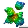
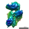
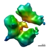
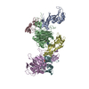
| Title | Reconfiguration of yeast 40S ribosomal subunit domains by the translation initiation multifactor complex. | |||||||||
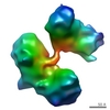
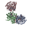
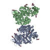
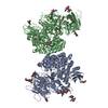
 Map data Map data | Reconstruction of the eukaryotic initiation factor multifactor complex (MFC). | |||||||||
 Sample Sample |
| |||||||||
| Biological species |   Saccharomyces cerevisiae (brewer's yeast) Saccharomyces cerevisiae (brewer's yeast) | |||||||||
| Method | single particle reconstruction / cryo EM / Resolution: 14.0 Å | |||||||||
 Authors Authors | Gilbert RJC / Gordiyenko Y / von der Haar T | |||||||||
 Citation Citation | Journal: Proc Natl Acad Sci U S A / Year: 2007 Title: Reconfiguration of yeast 40S ribosomal subunit domains by the translation initiation multifactor complex. Authors: Robert J C Gilbert / Yulya Gordiyenko / Tobias von der Haar / Andreas F-P Sonnen / Gregor Hofmann / Maria Nardelli / David I Stuart / John E G McCarthy /  Abstract: In the process of protein synthesis, the small (40S) subunit of the eukaryotic ribosome is recruited to the capped 5' end of the mRNA, from which point it scans along the 5' untranslated region in ...In the process of protein synthesis, the small (40S) subunit of the eukaryotic ribosome is recruited to the capped 5' end of the mRNA, from which point it scans along the 5' untranslated region in search of a start codon. However, the 40S subunit alone is not capable of functional association with cellular mRNA species; it has to be prepared for the recruitment and scanning steps by interactions with a group of eukaryotic initiation factors (eIFs). In budding yeast, an important subset of these factors (1, 2, 3, and 5) can form a multifactor complex (MFC). Here, we describe cryo-EM reconstructions of the 40S subunit, of the MFC, and of 40S complexes with MFC factors plus eIF1A. These studies reveal the positioning of the core MFC on the 40S subunit, and show how eIF-binding induces mobility in the head and platform and reconfigures the head-platform-body relationship. This is expected to increase the accessibility of the mRNA channel, thus enabling the 40S subunit to convert to a recruitment-competent state. | |||||||||
| History |
|
- Structure visualization
| Movie |
 Movie viewer Movie viewer |
|---|---|
| Structure viewer | EM map: SurfViewMolmilJmol/JSmol |
| Supplemental images |

- Downloads & links
-EMDB archive
| Map data | emd_1332.map.gz | 88.7 KB | EMDB map data format | |
|---|---|---|---|---|
| Header (meta data) | emd-1332-v30.xmlemd-1332.xml | 13.4 KB 13.4 KB | Display Display | EMDB header |
| Images |  1332.gif 1332.gif | 54 KB | ||
| Archive directory |  http://ftp.pdbj.org/pub/emdb/structures/EMD-1332ftp://ftp.pdbj.org/pub/emdb/structures/EMD-1332 http://ftp.pdbj.org/pub/emdb/structures/EMD-1332ftp://ftp.pdbj.org/pub/emdb/structures/EMD-1332 | HTTPS FTP |
-Related structure data


-Links
| EMDB pages | EMDB (EBI/PDBe) / EMDataResource |
|---|---|
| Related items in Molecule of the Month |

-Map
| File | Download / File: emd_1332.map.gz / Format: CCP4 / Size: 7.8 MB / Type: IMAGE STORED AS FLOATING POINT NUMBER (4 BYTES) | ||||||||||||||||||||||||||||||||||||||||||||||||||||||||||||||||||||
|---|---|---|---|---|---|---|---|---|---|---|---|---|---|---|---|---|---|---|---|---|---|---|---|---|---|---|---|---|---|---|---|---|---|---|---|---|---|---|---|---|---|---|---|---|---|---|---|---|---|---|---|---|---|---|---|---|---|---|---|---|---|---|---|---|---|---|---|---|---|
| Annotation | Reconstruction of the eukaryotic initiation factor multifactor complex (MFC). | ||||||||||||||||||||||||||||||||||||||||||||||||||||||||||||||||||||
| Projections & slices | Image control
Images are generated by Spider. | ||||||||||||||||||||||||||||||||||||||||||||||||||||||||||||||||||||
| Voxel size | X=Y=Z: 3.33 Å | ||||||||||||||||||||||||||||||||||||||||||||||||||||||||||||||||||||
| Density |
| ||||||||||||||||||||||||||||||||||||||||||||||||||||||||||||||||||||
| Symmetry | Space group: 1 | ||||||||||||||||||||||||||||||||||||||||||||||||||||||||||||||||||||
| Details | EMDB XML:
CCP4 map header:
| ||||||||||||||||||||||||||||||||||||||||||||||||||||||||||||||||||||
 Z (Sec.)
 X (Row.)
 Y (Col.)


-Supplemental data
- Sample components
-Entire : Eukaryotic translation multifactor complex
| Entire | Name: Eukaryotic translation multifactor complex |
|---|---|
| Components |
|
-Supramolecule #1000: Eukaryotic translation multifactor complex
| Supramolecule | Name: Eukaryotic translation multifactor complex / type: sample / ID: 1000 / Oligomeric state: Met-tRNA-eIF2-GMP-PNP-eIF3-eIF5 / Number unique components: 5 |
|---|---|
| Molecular weight | Theoretical: 1.954 MDa |
-Supramolecule #1: Small subunit
| Supramolecule | Name: Small subunit / type: complex / ID: 1 / Name.synonym: 40S / Details: S. cerevisiae / Recombinant expression: No / Ribosome-details: ribosome-eukaryote: SSU 40S |
|---|---|
| Source (natural) | Organism: Saccharomyces cerevisiae (brewer's yeast) / synonym: Baker's Yeast |
| Molecular weight | Experimental: 1.4 MDa / Theoretical: 1.4 MDa |
-Macromolecule #1: tRNA
| Macromolecule | Name: tRNA / type: rna / ID: 1 / Classification: TRANSFER / Structure: DOUBLE HELIX / Synthetic?: No |
|---|---|
| Source (natural) | Organism: Saccharomyces cerevisiae (brewer's yeast) / synonym: Baker's yeast |
| Molecular weight | Experimental: 23 KDa / Theoretical: 23 KDa |
-Macromolecule #2: eukaryotic translation initiation factor 2
| Macromolecule | Name: eukaryotic translation initiation factor 2 / type: protein_or_peptide / ID: 2 / Name.synonym: eIF2 / Number of copies: 1 / Oligomeric state: heterotrimer / Recombinant expression: Yes |
|---|---|
| Source (natural) | Organism: Saccharomyces cerevisiae (brewer's yeast) / synonym: Baker's yeast / Location in cell: Cytosol |
| Molecular weight | Experimental: 124 KDa / Theoretical: 124 KDa |
| Recombinant expression | Organism: Saccharomyces cerevisiae (brewer's yeast) |
-Macromolecule #3: eukaryotic translation initiation factor 3
| Macromolecule | Name: eukaryotic translation initiation factor 3 / type: protein_or_peptide / ID: 3 / Name.synonym: eIF3 / Number of copies: 1 / Oligomeric state: Heteropentamer / Recombinant expression: Yes |
|---|---|
| Source (natural) | Organism: Saccharomyces cerevisiae (brewer's yeast) / synonym: Baker's yeast / Location in cell: Cytosol |
| Molecular weight | Experimental: 362 KDa / Theoretical: 362 KDa |
| Recombinant expression | Organism: Saccharomyces cerevisiae (brewer's yeast) |
-Macromolecule #4: eukaryotic translation initiation factor 1
| Macromolecule | Name: eukaryotic translation initiation factor 1 / type: protein_or_peptide / ID: 4 / Name.synonym: eIF5 / Number of copies: 1 / Oligomeric state: Monomer / Recombinant expression: Yes |
|---|---|
| Source (natural) | Organism: Saccharomyces cerevisiae (brewer's yeast) / synonym: Baker's yeast / Location in cell: Cytosol |
| Molecular weight | Experimental: 45 KDa / Theoretical: 45 KDa |
| Recombinant expression | Organism: Saccharomyces cerevisiae (brewer's yeast) |
-Experimental details
-Structure determination
| Method | cryo EM |
|---|---|
 Processing Processing | single particle reconstruction |
| Aggregation state | particle |
-Sample preparation
| Buffer | pH: 7.4 Details: 38mM HEPES, 135mM KAc, 3.25mM MgAc2, 5mM beta-mercaptoethanol, 10uM GMP-PNP |
|---|---|
| Grid | Details: 300 mesh copper grid with lacey carbon film |
| Vitrification | Cryogen name: ETHANE / Instrument: HOMEMADE PLUNGER / Details: Vitrification instrument: Home-made plunger Method: Blot with Whatman number 1 paper for 1-2 seconds prior to plunging. |
- Electron microscopy
| Microscope | FEI/PHILIPS CM200FEG |
|---|---|
| Electron beam | Acceleration voltage: 200 kV / Electron source: FIELD EMISSION GUN |
| Electron optics | Illumination mode: FLOOD BEAM / Imaging mode: BRIGHT FIELDBright-field microscopy / Cs: 2 mm / Nominal defocus max: 11.685 µm / Nominal defocus min: 2.055 µm / Nominal magnification: 50000 |
| Sample stage | Specimen holder: Eucentric / Specimen holder model: GATAN LIQUID NITROGEN |
| Temperature | Average: 100 K |
| Alignment procedure | Legacy - Astigmatism: Astigmatism corrected at 100,000 x |
| Image recording | Category: FILM / Film or detector model: KODAK SO-163 FILM / Digitization - Scanner: OTHER / Digitization - Sampling interval: 8.33 µm / Number real images: 30 / Bits/pixel: 8 |
-Image processing
| CTF correction | Details: Per micrograph |
|---|---|
| Final angle assignment | Details: SPIDER Euler angle convention |
| Final reconstruction | Applied symmetry - Point group: C1 (asymmetric) / Algorithm: OTHER / Resolution.type: BY AUTHOR / Resolution: 14.0 Å / Resolution method: FSC 0.5 CUT-OFF / Software - Name: IMAGIC, EMAN, FREALIGN, SPIDER, GAP Details: Final maps were computed from CTF-corrected (by phase flipping) images, and scaled in Fourier space to a scattering model of the structure. Number images used: 19946 |
| Details | The particles were selected manually. |
-Atomic model buiding 1
| Software | Name: GAP |
|---|---|
| Details | Protocol: Rigid body |
| Refinement | Space: REAL / Protocol: RIGID BODY FIT / Target criteria: Real space CC and R-factor |
