 Movie
Movie Controller

[English] 日本語
 Yorodumi
- EMDB-1326: Minute virus of mice, a parvovirus, in complex with the Fab fragm... -
+ Open data

- Basic information
| Entry | Database: EMDB / ID: EMD-1326 | |||||||||
|---|---|---|---|---|---|---|---|---|---|---|
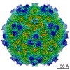
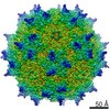
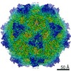
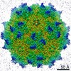
| Title | Minute virus of mice, a parvovirus, in complex with the Fab fragment of a neutralizing monoclonal antibody. | |||||||||
 Map data Map data | Minute | |||||||||
 Sample Sample |
| |||||||||
| Biological species |  | |||||||||
| Method | single particle reconstruction / cryo EM / Resolution: 7.0 Å | |||||||||
 Authors Authors | Kaufmann B / Lopez-Bueno A / Garcia-Mateu M / Chipman PR / Nelson CDS / Parrish CR / Almendral JM / Rossmann MG | |||||||||
 Citation Citation | Journal: J Virol / Year: 2007 Title: Minute virus of mice, a parvovirus, in complex with the Fab fragment of a neutralizing monoclonal antibody. Authors: Bärbel Kaufmann / Alberto López-Bueno / Mauricio G Mateu / Paul R Chipman / Christian D S Nelson / Colin R Parrish / José M Almendral / Michael G Rossmann /  Abstract: The structure of virus-like particles of the lymphotropic, immunosuppressive strain of minute virus of mice (MVMi) in complex with the neutralizing Fab fragment of the mouse monoclonal antibody (MAb) ...The structure of virus-like particles of the lymphotropic, immunosuppressive strain of minute virus of mice (MVMi) in complex with the neutralizing Fab fragment of the mouse monoclonal antibody (MAb) B7 was determined by cryo-electron microscopy to 7-A resolution. The Fab molecule recognizes a conformational epitope at the vertex of a three-fold protrusion on the viral surface, thereby simultaneously engaging three symmetry-related viral proteins in binding. The location of the epitope close to the three-fold axis is consistent with the previous analysis of MVMi mutants able to escape from the B7 antibody. The binding site close to the symmetry axes sterically forbids the binding of more than one Fab molecule per spike. MAb as well as the Fab molecules inhibits the binding of the minute virus of mice (MVM) to permissive cells but can also neutralize MVM postattachment. This finding suggests that the interaction of B7 with three symmetry-related viral subunits at each spike hinders structural transitions in the viral capsid essential during viral entry. | |||||||||
| History |
|
- Structure visualization
| Movie |
 Movie viewer Movie viewer |
|---|---|
| Structure viewer | EM map: SurfViewMolmilJmol/JSmol |
| Supplemental images |

- Downloads & links
-EMDB archive
| Map data | emd_1326.map.gz | 30.2 MB | EMDB map data format | |
|---|---|---|---|---|
| Header (meta data) | emd-1326-v30.xmlemd-1326.xml | 12.7 KB 12.7 KB | Display Display | EMDB header |
| Images |  1326.gif 1326.gif | 47.1 KB | ||
| Archive directory |  http://ftp.pdbj.org/pub/emdb/structures/EMD-1326ftp://ftp.pdbj.org/pub/emdb/structures/EMD-1326 http://ftp.pdbj.org/pub/emdb/structures/EMD-1326ftp://ftp.pdbj.org/pub/emdb/structures/EMD-1326 | HTTPS FTP |
-Validation report
| Summary document | emd_1326_validation.pdf.gz | 269.7 KB | Display | EMDB validaton report |
|---|---|---|---|---|
| Full document | emd_1326_full_validation.pdf.gz | 268.8 KB | Display | |
| Data in XML | emd_1326_validation.xml.gz | 6.5 KB | Display | |
| Arichive directory | https://ftp.pdbj.org/pub/emdb/validation_reports/EMD-1326ftp://ftp.pdbj.org/pub/emdb/validation_reports/EMD-1326 | HTTPS FTP |
-Related structure data
| Similar structure data |
|---|


-Links
| EMDB pages | EMDB (EBI/PDBe) / EMDataResource |
|---|
-Map
| File | Download / File: emd_1326.map.gz / Format: CCP4 / Size: 81.8 MB / Type: IMAGE STORED AS FLOATING POINT NUMBER (4 BYTES) | ||||||||||||||||||||||||||||||||||||||||||||||||||||||||||||||||||||
|---|---|---|---|---|---|---|---|---|---|---|---|---|---|---|---|---|---|---|---|---|---|---|---|---|---|---|---|---|---|---|---|---|---|---|---|---|---|---|---|---|---|---|---|---|---|---|---|---|---|---|---|---|---|---|---|---|---|---|---|---|---|---|---|---|---|---|---|---|---|
| Annotation | Minute | ||||||||||||||||||||||||||||||||||||||||||||||||||||||||||||||||||||
| Voxel size | X=Y=Z: 1.47371 Å | ||||||||||||||||||||||||||||||||||||||||||||||||||||||||||||||||||||
| Density |
| ||||||||||||||||||||||||||||||||||||||||||||||||||||||||||||||||||||
| Symmetry | Space group: 1 | ||||||||||||||||||||||||||||||||||||||||||||||||||||||||||||||||||||
| Details | EMDB XML:
CCP4 map header:
| ||||||||||||||||||||||||||||||||||||||||||||||||||||||||||||||||||||
-Supplemental data
- Sample components
-Entire : VLPs of Minute Virus of Mice strain i complexed with Fab fragment...
| Entire | Name: VLPs of Minute Virus of Mice strain i complexed with Fab fragment of neutralizing monoclonal antibody B7 |
|---|---|
| Components |
|
-Supramolecule #1000: VLPs of Minute Virus of Mice strain i complexed with Fab fragment...
| Supramolecule | Name: VLPs of Minute Virus of Mice strain i complexed with Fab fragment of neutralizing monoclonal antibody B7 type: sample / ID: 1000 Oligomeric state: twenty Fab fragments bind to one icosahedral virus particle Number unique components: 2 |
|---|---|
| Molecular weight | Theoretical: 4.84 MDa |
-Supramolecule #1: Minute Virus of Mice virus like particles
| Supramolecule | Name: Minute Virus of Mice virus like particles / type: virus / ID: 1 / Name.synonym: MVMi / Details: icosahedral particle / Sci species name: Minute Virus of Mice virus like particles / Virus type: VIRUS-LIKE PARTICLE / Virus isolate: STRAIN / Virus enveloped: No / Virus empty: Yes / Syn species name: MVMi |
|---|---|
| Host (natural) | synonym: VERTEBRATES |
| Molecular weight | Experimental: 3.8 MDa |
| Virus shell | Shell ID: 1 / Name: VP2 protein shell / Diameter: 280 Å / T number (triangulation number): 1 |
-Macromolecule #1: B7 Fab fragment
| Macromolecule | Name: B7 Fab fragment / type: protein_or_peptide / ID: 1 / Name.synonym: Fab B7 / Details: monomer / Oligomeric state: monomer / Recombinant expression: Yes |
|---|---|
| Source (natural) | Organism: |
| Molecular weight | Experimental: 50 KDa |
| Recombinant expression | Organism: hybridoma cell line (unknown) |
-Experimental details
-Structure determination
| Method | cryo EM |
|---|---|
 Processing Processing | single particle reconstruction |
| Aggregation state | particle |
-Sample preparation
| Buffer | pH: 7.5 / Details: 10mM Tris-HCl, 100mM NaCl |
|---|---|
| Grid | Details: 400 mesh copper |
| Vitrification | Cryogen name: ETHANE / Instrument: HOMEMADE PLUNGER Details: Vitrification instrument: guillotine-style plunge freezing device Method: A small vial of ethane is placed inside a larger liquid nitrogen reservoir. The grid holding a few microliters of the sample is held in place at the bottom of a plunger by the means of fine ...Method: A small vial of ethane is placed inside a larger liquid nitrogen reservoir. The grid holding a few microliters of the sample is held in place at the bottom of a plunger by the means of fine tweezers. Once the ethane in the vial is completely frozen, it needs to be slightly melted. When the liquid ethane is ready, a piece of filter paper is then pressed against the sample to blot of excess buffer, sufficient to leave a thin layer on the grid. After a predetermined time, the filter paper is removed, and the plunger is allowed to drop into the liquid ethane. Once the grid enters the liquid ethane, the sample is rapidly frozen, and the grid is transferred under liquid nitrogen to a storage box immersed liquid nitrogen for later use in the microscope. |
- Electron microscopy
| Microscope | FEI/PHILIPS CM300FEG/T |
|---|---|
| Temperature | Average: 98 K |
| Alignment procedure | Legacy - Astigmatism: live FFT at 200K |
| Details | low dose |
| Date | Oct 28, 2003 |
| Image recording | Category: FILM / Film or detector model: KODAK SO-163 FILM / Digitization - Scanner: ZEISS SCAI / Digitization - Sampling interval: 7 µm / Number real images: 14 / Average electron dose: 22 e/Å2 / Od range: 1.15 / Bits/pixel: 8 |
| Tilt angle min | 0 |
| Tilt angle max | 0 |
| Electron beam | Acceleration voltage: 300 kV / Electron source:  FIELD EMISSION GUN FIELD EMISSION GUN |
| Electron optics | Calibrated magnification: 47190 / Illumination mode: FLOOD BEAM / Imaging mode: BRIGHT FIELD / Cs: 2.0 mm / Nominal defocus max: 3.198 µm / Nominal defocus min: 2.052 µm / Nominal magnification: 45000 |
| Sample stage | Specimen holder: EUCENTRIC / Specimen holder model: GATAN LIQUID NITROGEN |
-Image processing
| CTF correction | Details: each particle |
|---|---|
| Final reconstruction | Applied symmetry - Point group: I (icosahedral) / Algorithm: OTHER / Resolution.type: BY AUTHOR / Resolution: 7.0 Å / Resolution method: FSC 0.5 CUT-OFF / Software - Name: EMPFT, POR, P3DR Details: final map includes data to 6.8 Ang resolution (fsc 0.3 cut-off); magnification of final map standardized to a map calculated from MVMi model coordinates (PDB accession no 1Z1C) resulting in ...Details: final map includes data to 6.8 Ang resolution (fsc 0.3 cut-off); magnification of final map standardized to a map calculated from MVMi model coordinates (PDB accession no 1Z1C) resulting in final pixel separation of 1.474Ang Number images used: 6375 |
-Atomic model buiding 1
| Software | Name: EMFIT SR5 |
|---|---|
| Details | Protocol: rigid body. The atomic coordinates of a Fab B7 homology model were fitted into difference map calculated between the MVM-Fab complex and MVM on its own. |
| Refinement | Space: REAL / Protocol: RIGID BODY FIT Target criteria: sum of density at each atomic positon, lack of atoms in negative density, distance restraints |
