 Movie
Movie Controller

[English] 日本語
 Yorodumi
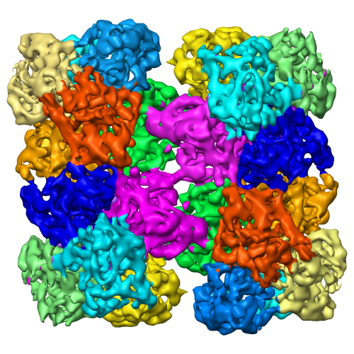
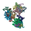
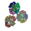
- EMDB-5101: 24-meric Scorpion Hemocyanin Activated State cryo-EM Density Map ... -
+ Open data

- Basic information
| Entry | Database: EMDB / ID: EMD-5101 | |||||||||
|---|---|---|---|---|---|---|---|---|---|---|
| Title | 24-meric Scorpion Hemocyanin Activated State cryo-EM Density Map at 8 Angstrom Resolution | |||||||||
 Map data Map data | This is the Hemocyanin activated cryo-EM density map | |||||||||
 Sample Sample |
| |||||||||
 Keywords Keywords |  Hemocyanin / Hc / Phenolxoidase activity / Tyrosinase (Ty) / Catecholoxidase (CO) / Enzyme / SDS / cryo-EM / single particle analysis Hemocyanin / Hc / Phenolxoidase activity / Tyrosinase (Ty) / Catecholoxidase (CO) / Enzyme / SDS / cryo-EM / single particle analysis | |||||||||
| Function / homology |  Function and homology informationchloride ion binding / oxygen carrier activity / oxidoreductase activity / copper ion binding / extracellular region Function and homology informationchloride ion binding / oxygen carrier activity / oxidoreductase activity / copper ion binding / extracellular regionSimilarity search - Function | |||||||||
| Biological species | unidentified (others) | |||||||||
| Method | single particle reconstruction / cryo EM / Resolution: 8.0 Å | |||||||||
 Authors Authors | Cong Y / Zhang Q / Woolford D / Schweikardt T / Khant H / Ludtke S / Chiu W / Decker H | |||||||||
 Citation Citation | Journal: Structure / Year: 2009 Title: Structural mechanism of SDS-induced enzyme activity of scorpion hemocyanin revealed by electron cryomicroscopy. Authors: Yao Cong / Qinfen Zhang / David Woolford / Thorsten Schweikardt / Htet Khant / Matthew Dougherty / Steven J Ludtke / Wah Chiu / Heinz Decker /  Abstract: Phenoloxidases (POs) occur in all organisms and are involved in skin and hair coloring in mammals, and initiating melanization in wound healing. Mutation or overexpression of PO can cause albinism or ...Phenoloxidases (POs) occur in all organisms and are involved in skin and hair coloring in mammals, and initiating melanization in wound healing. Mutation or overexpression of PO can cause albinism or melanoma, respectively. SDS can convert inactive PO and the oxygen carrier hemocyanin (Hc) into enzymatically active PO. Here we present single-particle cryo-EM maps at subnanometer resolution and pseudoatomic models of the 24-oligomeric Hc from scorpion Pandinus imperator in resting and SDS-activated states. Our structural analyses led to a plausible mechanism of Hc enzyme PO activation: upon SDS activation, the intrinsically flexible Hc domain I twists away from domains II and III in each subunit, exposing the entrance to the active site; this movement is stabilized by enhanced interhexamer and interdodecamer interactions, particularly in the central linker subunits. This mechanism could be applicable to other type 3 copper proteins, as the active site is highly conserved. | |||||||||
| History |
|
- Structure visualization
| Movie |
Movie viewer |
|---|---|
| Structure viewer | EM map: SurfViewMolmilJmol/JSmol |
| Supplemental images |
- Downloads & links
-EMDB archive
| Map data | emd_5101.map.gz | 31.9 MB | EMDB map data format | |
|---|---|---|---|---|
| Header (meta data) | emd-5101-v30.xmlemd-5101.xml | 11.4 KB 11.4 KB | Display Display | EMDB header |
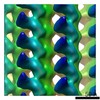
| Images |  emd_5101_1.png emd_5101_1.png | 353.7 KB | ||
| Archive directory |  http://ftp.pdbj.org/pub/emdb/structures/EMD-5101ftp://ftp.pdbj.org/pub/emdb/structures/EMD-5101 http://ftp.pdbj.org/pub/emdb/structures/EMD-5101ftp://ftp.pdbj.org/pub/emdb/structures/EMD-5101 | HTTPS FTP |
-Related structure data
| Related structure data |  3ixwMC  5100C 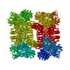 3ixvC M: atomic model generated by this map C: citing same article ( |
|---|---|
| Similar structure data |
-Links
| EMDB pages | EMDB (EBI/PDBe) / EMDataResource |
|---|
-Map
| File | Download / File: emd_5101.map.gz / Format: CCP4 / Size: 33.5 MB / Type: IMAGE STORED AS FLOATING POINT NUMBER (4 BYTES) | ||||||||||||||||||||||||||||||||||||||||||||||||||||||||||||||||||||
|---|---|---|---|---|---|---|---|---|---|---|---|---|---|---|---|---|---|---|---|---|---|---|---|---|---|---|---|---|---|---|---|---|---|---|---|---|---|---|---|---|---|---|---|---|---|---|---|---|---|---|---|---|---|---|---|---|---|---|---|---|---|---|---|---|---|---|---|---|---|
| Annotation | This is the Hemocyanin activated cryo-EM density map | ||||||||||||||||||||||||||||||||||||||||||||||||||||||||||||||||||||
| Voxel size | X=Y=Z: 1.8 Å | ||||||||||||||||||||||||||||||||||||||||||||||||||||||||||||||||||||
| Density |
| ||||||||||||||||||||||||||||||||||||||||||||||||||||||||||||||||||||
| Symmetry | Space group: 1 | ||||||||||||||||||||||||||||||||||||||||||||||||||||||||||||||||||||
| Details | EMDB XML:
CCP4 map header:
| ||||||||||||||||||||||||||||||||||||||||||||||||||||||||||||||||||||
-Supplemental data
- Sample components
-Entire : Hemocyanin from scorpion Pandinus imperator (Activated State)
| Entire | Name: Hemocyanin from scorpion Pandinus imperator (Activated State) |
|---|---|
| Components |
|
-Supramolecule #1000: Hemocyanin from scorpion Pandinus imperator (Activated State)
| Supramolecule | Name: Hemocyanin from scorpion Pandinus imperator (Activated State) type: sample / ID: 1000 / Details: treated by 2mM SDS to activate Hc / Oligomeric state: 24-mer / Number unique components: 1 |
|---|---|
| Molecular weight | Experimental: 1.7 MDa / Theoretical: 1.7 MDa / Method: Sedimentation |
-Macromolecule #1: Pandinus imperator
| Macromolecule | Name: Pandinus imperator / type: protein_or_peptide / ID: 1 / Name.synonym: scorpion / Number of copies: 1 / Oligomeric state: 24-mer / Recombinant expression: No / Database: NCBI |
|---|---|
| Source (natural) | Organism: unidentified (others) / Location in cell: hemolymph |
| Molecular weight | Experimental: 1.7 MDa / Theoretical: 1.7 MDa |
| Sequence | GO: oxygen carrier activity / InterPro: Hemocyanin/hexamerin middle domain |
-Experimental details
-Structure determination
| Method | cryo EM |
|---|---|
 Processing Processing | single particle reconstruction |
| Aggregation state | particle |
-Sample preparation
| Concentration | 0.5 mg/mL |
|---|---|
| Buffer | pH: 7.8 Details: 100 mM TRIS/HCL at pH 7.8, 10 mM CaCl2 and 10 mM MgCl2 |
| Grid | Details: 200 mesh copper grid |
| Vitrification | Cryogen name: ETHANE / Chamber humidity: 95 % / Chamber temperature: 101 K / Instrument: OTHER / Details: Vitrification instrument: FEI vitrobot / Method: two side blotting for 1 second before plunging |
- Electron microscopy
| Microscope | JEOL 3200FSC |
|---|---|
| Electron beam | Acceleration voltage: 300 kV / Electron source: FIELD EMISSION GUN |
| Electron optics | Illumination mode: FLOOD BEAM / Imaging mode: BRIGHT FIELDBright-field microscopy / Cs: 4.1 mm / Nominal defocus max: 3.5 µm / Nominal defocus min: 1.0 µm / Nominal magnification: 50000 |
| Specialist optics | Energy filter - Name: in-column omega energy filter / Energy filter - Lower energy threshold: 0.0 eV / Energy filter - Upper energy threshold: 20.0 eV |
| Sample stage | Specimen holder: Side Entry / Specimen holder model: JEOL 3200FSC CRYOHOLDER |
| Temperature | Min: 101 K / Max: 101.2 K / Average: 101 K |
| Alignment procedure | Legacy - Astigmatism: objective lens astigmatism correction |
| Details | JEOL 3200FSC MDS low dose method |
| Date | May 31, 2007 |
| Image recording | Category: FILM / Film or detector model: KODAK SO-163 FILM / Digitization - Scanner: NIKON SUPER COOLSCAN 9000 / Digitization - Sampling interval: 6.35 µm / Number real images: 200 / Average electron dose: 18 e/Å2 |
| Tilt angle min | 0 |
| Tilt angle max | 0 |
-Image processing
| CTF correction | Details: each micrograph |
|---|---|
| Final reconstruction | Algorithm: OTHER / Resolution.type: BY AUTHOR / Resolution: 8.0 Å / Resolution method: FSC 0.5 CUT-OFF / Software - Name: EMAN Details: Final refinement using FRM2D (Fast Rotational Matching) image alignment method Number images used: 13400 |
