 Movie
Movie Controller


+ Open data

- Basic information
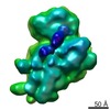
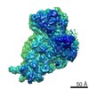
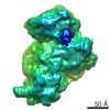
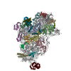
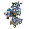
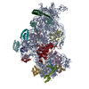
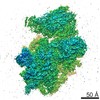
| Entry | Database: EMDB / ID: EMD-1770 | |||||||||
|---|---|---|---|---|---|---|---|---|---|---|
| Title | 30S-002mRNA (after classification) | |||||||||
 Map data Map data | 30S-002mRNA (after classification) | |||||||||
 Sample Sample |
| |||||||||
| Biological species |  | |||||||||
| Method | single particle reconstruction / cryo EM / negative staining / Resolution: 21.0 Å | |||||||||
 Authors Authors | Julian P / Milon P / Agirrezabala X / Lasso G / Gil D / Rodnina MV / Valle M | |||||||||
 Citation Citation | Journal: PLoS Biol / Year: 2011 Title: The Cryo-EM structure of a complete 30S translation initiation complex from Escherichia coli. Authors: Patricia Julián / Pohl Milon / Xabier Agirrezabala / Gorka Lasso / David Gil / Marina V Rodnina / Mikel Valle /  Abstract: Formation of the 30S initiation complex (30S IC) is an important checkpoint in regulation of gene expression. The selection of mRNA, correct start codon, and the initiator fMet-tRNA(fMet) requires ...Formation of the 30S initiation complex (30S IC) is an important checkpoint in regulation of gene expression. The selection of mRNA, correct start codon, and the initiator fMet-tRNA(fMet) requires the presence of three initiation factors (IF1, IF2, IF3) of which IF3 and IF1 control the fidelity of the process, while IF2 recruits fMet-tRNA(fMet). Here we present a cryo-EM reconstruction of the complete 30S IC, containing mRNA, fMet-tRNA(fMet), IF1, IF2, and IF3. In the 30S IC, IF2 contacts IF1, the 30S subunit shoulder, and the CCA end of fMet-tRNA(fMet), which occupies a novel P/I position (P/I1). The N-terminal domain of IF3 contacts the tRNA, whereas the C-terminal domain is bound to the platform of the 30S subunit. Binding of initiation factors and fMet-tRNA(fMet) induces a rotation of the head relative to the body of the 30S subunit, which is likely to prevail through 50S subunit joining until GTP hydrolysis and dissociation of IF2 take place. The structure provides insights into the mechanism of mRNA selection during translation initiation. | |||||||||
| History |
|
- Structure visualization
| Movie |
 Movie viewer Movie viewer |
|---|---|
| Structure viewer | EM map: SurfViewMolmilJmol/JSmol |
| Supplemental images |

- Downloads & links
-EMDB archive
| Map data | emd_1770.map.gz | 7.8 MB | EMDB map data format | |
|---|---|---|---|---|
| Header (meta data) | emd-1770-v30.xmlemd-1770.xml | 7.8 KB 7.8 KB | Display Display | EMDB header |
| Images | EMD-1770.tif | 3.4 MB | ||
| Archive directory |  http://ftp.pdbj.org/pub/emdb/structures/EMD-1770ftp://ftp.pdbj.org/pub/emdb/structures/EMD-1770 http://ftp.pdbj.org/pub/emdb/structures/EMD-1770ftp://ftp.pdbj.org/pub/emdb/structures/EMD-1770 | HTTPS FTP |
-Validation report
| Summary document | emd_1770_validation.pdf.gz | 208.1 KB | Display | EMDB validaton report |
|---|---|---|---|---|
| Full document | emd_1770_full_validation.pdf.gz | 207.2 KB | Display | |
| Data in XML | emd_1770_validation.xml.gz | 5.2 KB | Display | |
| Arichive directory | https://ftp.pdbj.org/pub/emdb/validation_reports/EMD-1770ftp://ftp.pdbj.org/pub/emdb/validation_reports/EMD-1770 | HTTPS FTP |
-Related structure data
-Links
| EMDB pages | EMDB (EBI/PDBe) / EMDataResource |
|---|---|
| Related items in Molecule of the Month |

-Map
| File | Download / File: emd_1770.map.gz / Format: CCP4 / Size: 8.2 MB / Type: IMAGE STORED AS FLOATING POINT NUMBER (4 BYTES) | ||||||||||||||||||||||||||||||||||||||||||||||||||||||||||||||||||||
|---|---|---|---|---|---|---|---|---|---|---|---|---|---|---|---|---|---|---|---|---|---|---|---|---|---|---|---|---|---|---|---|---|---|---|---|---|---|---|---|---|---|---|---|---|---|---|---|---|---|---|---|---|---|---|---|---|---|---|---|---|---|---|---|---|---|---|---|---|---|
| Annotation | 30S-002mRNA (after classification) | ||||||||||||||||||||||||||||||||||||||||||||||||||||||||||||||||||||
| Voxel size | X=Y=Z: 2.8 Å | ||||||||||||||||||||||||||||||||||||||||||||||||||||||||||||||||||||
| Density |
| ||||||||||||||||||||||||||||||||||||||||||||||||||||||||||||||||||||
| Symmetry | Space group: 1 | ||||||||||||||||||||||||||||||||||||||||||||||||||||||||||||||||||||
| Details | EMDB XML:
CCP4 map header:
| ||||||||||||||||||||||||||||||||||||||||||||||||||||||||||||||||||||
-Supplemental data
- Sample components
-Entire : 30S.mRNA complex
| Entire | Name: 30S.mRNA complex |
|---|---|
| Components |
|
-Supramolecule #1000: 30S.mRNA complex
| Supramolecule | Name: 30S.mRNA complex / type: sample / ID: 1000 / Number unique components: 2 |
|---|
-Supramolecule #1: Ribosome
| Supramolecule | Name: Ribosome / type: complex / ID: 1 / Recombinant expression: No / Ribosome-details: ribosome-prokaryote: SSU 30S |
|---|---|
| Source (natural) | Organism: |
-Macromolecule #1: mRNA
| Macromolecule | Name: mRNA / type: rna / ID: 1 / Classification: OTHER / Structure: SINGLE STRANDED / Synthetic?: No |
|---|---|
| Source (natural) | Organism: |
-Experimental details
-Structure determination
| Method | negative staining, cryo EM |
|---|---|
 Processing Processing | single particle reconstruction |
| Aggregation state | particle |
-Sample preparation
| Buffer | Details: 50 mM Tris-HCl, pH 7.5, 70 mM NH4Cl, 30 mM KCl, and 7 mM MgCl2 |
|---|---|
| Staining | Type: NEGATIVE / Details: Cryo_EM |
| Grid | Details: Quantifoil grids |
| Vitrification | Cryogen name: ETHANE / Chamber humidity: 100 % / Instrument: OTHER / Details: Vitrification instrument: Vitrobot |
- Electron microscopy
| Microscope | JEOL 2200FS |
|---|---|
| Image recording | Digitization - Scanner: ZEISS SCAI / Digitization - Sampling interval: 14 µm |
| Electron beam | Acceleration voltage: 200 kV / Electron source:  FIELD EMISSION GUN FIELD EMISSION GUN |
| Electron optics | Illumination mode: FLOOD BEAM / Imaging mode: BRIGHT FIELD / Nominal magnification: 50000 |
| Sample stage | Specimen holder: Cryo Specimen Holder / Specimen holder model: GATAN LIQUID NITROGEN |
-Image processing
| Final reconstruction | Applied symmetry - Point group: C1 (asymmetric) / Resolution.type: BY AUTHOR / Resolution: 21.0 Å / Resolution method: FSC 0.5 CUT-OFF / Software - Name: Spire / Number images used: 48000 |
|---|---|
| Final two d classification | Number classes: 2 |
