 Movie
Movie Controller

[English] 日本語
 Yorodumi
- EMDB-1698: Three-dimensional structure of TspO by electron cryo-microscopy o... -
+ Open data

- Basic information
| Entry | Database: EMDB / ID: EMD-1698 | |||||||||
|---|---|---|---|---|---|---|---|---|---|---|
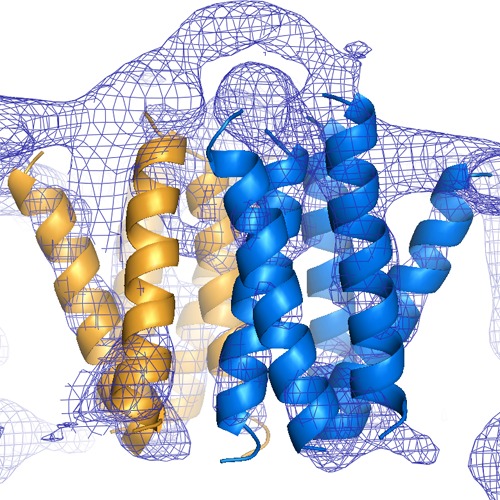
| Title | Three-dimensional structure of TspO by electron cryo-microscopy of helical crystals. | |||||||||
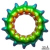
 Map data Map data | The map contains 120 Angstrom thick slab through the tube reconstruction. | |||||||||
 Sample Sample |
| |||||||||
 Keywords Keywords | TSPO / PBR / membrane protein / helical crystal | |||||||||
| Biological species |  Rhodobacter sphaeroides (bacteria) Rhodobacter sphaeroides (bacteria) | |||||||||
| Method | helical reconstruction / cryo EM / Resolution: 10.2 Å | |||||||||
 Authors Authors | Korkhov VM / Sachse C / Short JM / Tate CG | |||||||||
 Citation Citation | Journal: Structure / Year: 2010 Title: Three-dimensional structure of TspO by electron cryomicroscopy of helical crystals. Authors: Vladimir M Korkhov / Carsten Sachse / Judith M Short / Christopher G Tate /  Abstract: The 18 kDa TSPO protein is a polytopic mitochondrial outer membrane protein involved in a wide range of physiological functions and pathologies, including neurodegeneration and cancer. The ...The 18 kDa TSPO protein is a polytopic mitochondrial outer membrane protein involved in a wide range of physiological functions and pathologies, including neurodegeneration and cancer. The pharmacology of TSPO has been extensively studied, but little is known about its biochemistry, oligomeric state, and structure. We have expressed, purified, and characterized a homologous protein, TspO from Rhodobacter sphaeroides, and reconstituted it as helical crystals. Using electron cryomicroscopy and single-particle helical reconstruction, we have determined a three-dimensional structure of TspO at 10 A resolution. The structure suggests that monomeric TspO comprises five transmembrane alpha helices that form a homodimer, which is consistent with the dimeric state observed in detergent solution. Furthermore, the arrangement of transmembrane domains of individual TspO subunits indicates a possibility of two substrate translocation pathways per dimer. The structure provides the first insight into the molecular architecture of TSPO/PBR protein family that will serve as a framework for future studies. | |||||||||
| History |
|
- Structure visualization
| Movie |
 Movie viewer Movie viewer |
|---|---|
| Structure viewer | EM map: SurfViewMolmilJmol/JSmol |
| Supplemental images |
- Downloads & links
-EMDB archive
| Map data | emd_1698.map.gz | 21.8 MB | EMDB map data format | |
|---|---|---|---|---|
| Header (meta data) | emd-1698-v30.xmlemd-1698.xml | 10.8 KB 10.8 KB | Display Display | EMDB header |
| Images | 1698.tif | 909.4 KB | ||
| Archive directory |  http://ftp.pdbj.org/pub/emdb/structures/EMD-1698ftp://ftp.pdbj.org/pub/emdb/structures/EMD-1698 http://ftp.pdbj.org/pub/emdb/structures/EMD-1698ftp://ftp.pdbj.org/pub/emdb/structures/EMD-1698 | HTTPS FTP |
-Validation report
| Summary document | emd_1698_validation.pdf.gz | 227.3 KB | Display | EMDB validaton report |
|---|---|---|---|---|
| Full document | emd_1698_full_validation.pdf.gz | 226.4 KB | Display | |
| Data in XML | emd_1698_validation.xml.gz | 4.4 KB | Display | |
| Arichive directory | https://ftp.pdbj.org/pub/emdb/validation_reports/EMD-1698ftp://ftp.pdbj.org/pub/emdb/validation_reports/EMD-1698 | HTTPS FTP |
-Related structure data
| Similar structure data |
|---|

-Links
| EMDB pages | EMDB (EBI/PDBe) / EMDataResource |
|---|
-Map
| File | Download / File: emd_1698.map.gz / Format: CCP4 / Size: 23.3 MB / Type: IMAGE STORED AS FLOATING POINT NUMBER (4 BYTES) | ||||||||||||||||||||||||||||||||||||||||||||||||||||||||||||
|---|---|---|---|---|---|---|---|---|---|---|---|---|---|---|---|---|---|---|---|---|---|---|---|---|---|---|---|---|---|---|---|---|---|---|---|---|---|---|---|---|---|---|---|---|---|---|---|---|---|---|---|---|---|---|---|---|---|---|---|---|---|
| Annotation | The map contains 120 Angstrom thick slab through the tube reconstruction. | ||||||||||||||||||||||||||||||||||||||||||||||||||||||||||||
| Voxel size | X=Y=Z: 1.2 Å | ||||||||||||||||||||||||||||||||||||||||||||||||||||||||||||
| Density |
| ||||||||||||||||||||||||||||||||||||||||||||||||||||||||||||
| Symmetry | Space group: 1 | ||||||||||||||||||||||||||||||||||||||||||||||||||||||||||||
| Details | EMDB XML:
CCP4 map header:
| ||||||||||||||||||||||||||||||||||||||||||||||||||||||||||||
-Supplemental data
- Sample components
-Entire : TspO and Escherichia coli polar lipid extract
| Entire | Name: TspO and Escherichia coli polar lipid extract |
|---|---|
| Components |
|
-Supramolecule #1000: TspO and Escherichia coli polar lipid extract
| Supramolecule | Name: TspO and Escherichia coli polar lipid extract / type: sample / ID: 1000 / Oligomeric state: Helical / Number unique components: 2 |
|---|---|
| Molecular weight | Experimental: 13.5 MDa / Theoretical: 13.5 MDa |
-Supramolecule #1: Lipid extract
| Supramolecule | Name: Lipid extract / type: organelle_or_cellular_component / ID: 1 / Name.synonym: Lipid extract / Recombinant expression: No / Database: NCBI |
|---|---|
| Source (natural) | Organism: |
-Macromolecule #1: TspO
| Macromolecule | Name: TspO / type: protein_or_peptide / ID: 1 / Name.synonym: TspO / Oligomeric state: Helical / Recombinant expression: Yes |
|---|---|
| Source (natural) | Organism: Rhodobacter sphaeroides (bacteria) / synonym: Rhodobacter sphaeroides / Location in cell: Outer membrane |
| Molecular weight | Experimental: 18 KDa / Theoretical: 18 KDa |
| Recombinant expression | Organism: |
-Experimental details
-Structure determination
| Method | cryo EM |
|---|---|
 Processing Processing | helical reconstruction |
| Aggregation state | helical array |
-Sample preparation
| Concentration | 0.5 mg/mL |
|---|---|
| Buffer | pH: 7.5 / Details: 20 mM Tris, 100 mM NaCl, 2 mM EDTA |
| Vitrification | Cryogen name: ETHANE / Chamber temperature: 77.2 K / Instrument: HOMEMADE PLUNGER / Details: Vitrification instrument: Home built / Method: Back-side blotting |
| Details | Incubated and dialysed for 3 days with Escherichia coli polar lipid extract |
- Electron microscopy
| Microscope | FEI TECNAI F20 |
|---|---|
| Alignment procedure | Legacy - Astigmatism: Objective lens astigmatism was corrected at 100,000 times magnification |
| Date | May 30, 2007 |
| Image recording | Category: FILM / Film or detector model: KODAK SO-163 FILM / Digitization - Scanner: OTHER / Digitization - Sampling interval: 6 µm / Number real images: 36 / Average electron dose: 15 e/Å2 / Bits/pixel: 8 |
| Tilt angle min | 0 |
| Tilt angle max | 0 |
| Electron beam | Acceleration voltage: 200 kV / Electron source:  FIELD EMISSION GUN FIELD EMISSION GUN |
| Electron optics | Illumination mode: FLOOD BEAM / Imaging mode: BRIGHT FIELD / Cs: 2 mm / Nominal defocus max: 2.163 µm / Nominal defocus min: 0.8 µm / Nominal magnification: 50000 |
| Sample stage | Specimen holder: Side entry liquid nitrogen-cooled cryo specimen holder Specimen holder model: GATAN LIQUID NITROGEN |
| Experimental equipment |  Model: Tecnai F20 / Image courtesy: FEI Company |
-Image processing
| Details | 2 TspO molecules within dimer are related by an additional 2-fold axis parallel to tube axis. |
|---|---|
| Final reconstruction | Applied symmetry - Helical parameters - Δz: 32 Å Applied symmetry - Helical parameters - Δ&Phi: 9.6 ° Applied symmetry - Helical parameters - Axial symmetry: D12 (2x12 fold dihedral) Algorithm: OTHER / Resolution.type: BY AUTHOR / Resolution: 10.2 Å / Resolution method: FSC 0.5 CUT-OFF / Software - Name: SPIDER / Details: Imposition of 12-fold rotational symmetry |
| CTF correction | Details: Segment-specific CTF |
| Final angle assignment | Details: 2 degree increments, 0-360 degrees around helical axis, up to 12 degrees out-of-plane tilt |
