 Movie
Movie Controller

[English] 日本語
 Yorodumi
- EMDB-1422: Structure and composition of the Shigella flexneri "needle comple... -
+ Open data

- Basic information
| Entry | Database: EMDB / ID: EMD-1422 | |||||||||
|---|---|---|---|---|---|---|---|---|---|---|
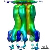
| Title | Structure and composition of the Shigella flexneri "needle complex", a part of its type III secreton. | |||||||||
 Map data Map data | Basal Body of Shigella flexneri T3SS - needle removed | |||||||||
 Sample Sample |
| |||||||||
| Function / homology | : / protein binding / Type III secretion system outer membrane pore YscC/HrcC / Type III secretion system, needle protein / Flagellar M-ring , N-terminal Function and homology information Function and homology information | |||||||||
| Biological species |  Shigella flexneri (bacteria) Shigella flexneri (bacteria) | |||||||||
| Method | single particle reconstruction / cryo EM / negative staining / Resolution: 17.0 Å | |||||||||
 Authors Authors | Blocker AJ / Jouihri N / Larquet E / Gounon P / Ebel F / Parsot C / Sansonetti P / Allaoui A | |||||||||
 Citation Citation | Journal: Mol Microbiol / Year: 2001 Title: Structure and composition of the Shigella flexneri "needle complex", a part of its type III secreton. Authors: A Blocker / N Jouihri / E Larquet / P Gounon / F Ebel / C Parsot / P Sansonetti / A Allaoui /  Abstract: Type III secretion systems (TTSSs or secretons), essential virulence determinants of many Gram-negative bacteria, serve to translocate proteins directly from the bacteria into the host cytoplasm. ...Type III secretion systems (TTSSs or secretons), essential virulence determinants of many Gram-negative bacteria, serve to translocate proteins directly from the bacteria into the host cytoplasm. Electron microscopy (EM) indicates that the TTSSs of Shigella flexneri are composed of: (1) an external needle; (2) a transmembrane domain; and (3) a cytoplasmic bulb. EM analysis of purified and negatively stained parts 1, 2 and a portion of 3 of the TTSS, together termed the "needle complex" (NC), produced an average image at 17 A resolution in which a base, an outer ring and a needle, inserted through the ring into the base, could be discerned. This analysis and cryoEM images of NCs indicated that the needle and base contain a central 2-3 nm canal. Five major NC components, MxiD, MxiG, MxiJ, MxiH and MxiI, were identified by N-terminal sequencing. MxiG and MxiJ are predicted to be inner membrane proteins and presumably form the base. MxiD is predicted to be an outer membrane protein and to form the outer ring. MxiH and MxiI are small hydrophilic proteins. Mutants lacking either of these proteins formed needleless secretons and were unable to secrete Ipa proteins. As MxiH was present in NCs in large molar excess, we propose that it is the major needle component. MxiI may cap at the external needle tip. | |||||||||
| History |
|
- Structure visualization
| Movie |
Movie viewer |
|---|---|
| Structure viewer | EM map: SurfViewMolmilJmol/JSmol |
| Supplemental images |
 UCSF Chimera
- Downloads & links
-EMDB archive
| Map data | emd_1422.map.gz | 3.3 MB | EMDB map data format | |
|---|---|---|---|---|
| Header (meta data) | emd-1422-v30.xmlemd-1422.xml | 14.9 KB 14.9 KB | Display Display | EMDB header |
| Images | 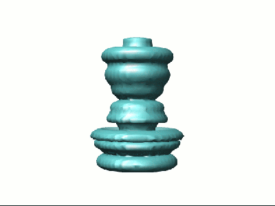 1422.gif 1422.gif | 17.5 KB | ||
| Archive directory |  http://ftp.pdbj.org/pub/emdb/structures/EMD-1422ftp://ftp.pdbj.org/pub/emdb/structures/EMD-1422 http://ftp.pdbj.org/pub/emdb/structures/EMD-1422ftp://ftp.pdbj.org/pub/emdb/structures/EMD-1422 | HTTPS FTP |
-Validation report
| Summary document | emd_1422_validation.pdf.gz | 199.4 KB | Display | EMDB validaton report |
|---|---|---|---|---|
| Full document | emd_1422_full_validation.pdf.gz | 198.5 KB | Display | |
| Data in XML | emd_1422_validation.xml.gz | 7 KB | Display | |
| Arichive directory | https://ftp.pdbj.org/pub/emdb/validation_reports/EMD-1422ftp://ftp.pdbj.org/pub/emdb/validation_reports/EMD-1422 | HTTPS FTP |
-Related structure data
| Similar structure data |
|---|
-Links
| EMDB pages | EMDB (EBI/PDBe) / EMDataResource |
|---|
-Map
| File | Download / File: emd_1422.map.gz / Format: CCP4 / Size: 100.6 MB / Type: IMAGE STORED AS FLOATING POINT NUMBER (4 BYTES) | ||||||||||||||||||||||||||||||||||||||||||||||||||||||||||||||||||||
|---|---|---|---|---|---|---|---|---|---|---|---|---|---|---|---|---|---|---|---|---|---|---|---|---|---|---|---|---|---|---|---|---|---|---|---|---|---|---|---|---|---|---|---|---|---|---|---|---|---|---|---|---|---|---|---|---|---|---|---|---|---|---|---|---|---|---|---|---|---|
| Annotation | Basal Body of Shigella flexneri T3SS - needle removed | ||||||||||||||||||||||||||||||||||||||||||||||||||||||||||||||||||||
| Voxel size | X=Y=Z: 2.2 Å | ||||||||||||||||||||||||||||||||||||||||||||||||||||||||||||||||||||
| Density |
| ||||||||||||||||||||||||||||||||||||||||||||||||||||||||||||||||||||
| Symmetry | Space group: 1 | ||||||||||||||||||||||||||||||||||||||||||||||||||||||||||||||||||||
| Details | EMDB XML:
CCP4 map header:
| ||||||||||||||||||||||||||||||||||||||||||||||||||||||||||||||||||||
-Supplemental data
- Sample components
-Entire : needle complex or basal body of the Shigella flexneri T3SS
| Entire | Name: needle complex or basal body of the Shigella flexneri T3SS |
|---|---|
| Components |
|
-Supramolecule #1000: needle complex or basal body of the Shigella flexneri T3SS
| Supramolecule | Name: needle complex or basal body of the Shigella flexneri T3SS type: sample / ID: 1000 / Oligomeric state: not yet fully determined / Number unique components: 5 |
|---|
-Macromolecule #1: MxiH
| Macromolecule | Name: MxiH / type: protein_or_peptide / ID: 1 / Details: none; copy number is approximative / Number of copies: 120 / Oligomeric state: helical polymer / Recombinant expression: No / Database: NCBI |
|---|---|
| Source (natural) | Organism: Shigella flexneri (bacteria) / Strain: M90T / Tissue: bacterium / Organelle: T3SS / Location in cell: extracellular |
| Molecular weight | Experimental: 9.265 MDa / Theoretical: 9.265 MDa |
| Sequence | InterPro: Type III secretion system, needle protein |
-Macromolecule #2: MxiI
| Macromolecule | Name: MxiI / type: protein_or_peptide / ID: 2 / Details: none; Copy number is approximative / Number of copies: 20 / Oligomeric state: probably helical / Recombinant expression: No / Database: NCBI |
|---|---|
| Source (natural) | Organism: Shigella flexneri (bacteria) / Strain: M90T / Cell: bacterium / Organelle: T3SS / Location in cell: periplasmic |
| Molecular weight | Experimental: 10.633 MDa / Theoretical: 10.633 MDa |
-Macromolecule #3: MxiD
| Macromolecule | Name: MxiD / type: protein_or_peptide / ID: 3 Details: experimental weight is theoretical weight, with predicted signal sequence removed; oligomeric state currently unknown Oligomeric state: oligomer / Recombinant expression: No / Database: NCBI |
|---|---|
| Source (natural) | Organism: Shigella flexneri (bacteria) / Strain: M90T / Cell: bacterium / Organelle: T3SS / Location in cell: outer membrane |
| Molecular weight | Experimental: 63.218 MDa / Theoretical: 60.749 MDa |
| Sequence | GO: GO: 0015448 InterPro: Type III secretion system outer membrane pore YscC/HrcC |
-Macromolecule #4: MxiG
| Macromolecule | Name: MxiG / type: protein_or_peptide / ID: 4 Details: no signal sequence cleavage in this protein; oligomeric state unknown Oligomeric state: oligomer / Recombinant expression: No / Database: NCBI |
|---|---|
| Source (natural) | Organism: Shigella flexneri (bacteria) / Strain: M90T / Cell: bacterium / Organelle: T3SS / Location in cell: inner membrane |
| Molecular weight | Experimental: 43.002 MDa / Theoretical: 43.002 MDa |
| Sequence | GO: protein binding |
-Macromolecule #5: MxiJ
| Macromolecule | Name: MxiJ / type: protein_or_peptide / ID: 5 Details: experimental weight is theoretical weight, with predicted signal sequence removed; oligomeric state unknown Oligomeric state: oligomer / Recombinant expression: No / Database: NCBI |
|---|---|
| Source (natural) | Organism: Shigella flexneri (bacteria) / Strain: M90T / Cell: bacterium / Organelle: T3SS / Location in cell: inner membrane |
| Molecular weight | Experimental: 27.509 MDa / Theoretical: 25.488 MDa |
| Sequence | GO: protein binding / InterPro: Flagellar M-ring , N-terminal |
-Experimental details
-Structure determination
| Method | negative staining, cryo EM |
|---|---|
 Processing Processing | single particle reconstruction |
| Aggregation state | particle |
-Sample preparation
| Buffer | pH: 7.4 Details: samples made in 50 mM Tris pH 8, 5 mM EDTA, 0.1 % v/v Triton X-100 and diluted 1:5 in 20 mM Tris pH 7.4 |
|---|---|
| Staining | Type: NEGATIVE / Details: 1% uranyl acetate pH 7.5 |
| Grid | Details: carbon-coated glow-discharged copper grids |
| Vitrification | Cryogen name: ETHANE |
- Electron microscopy
| Microscope | FEI/PHILIPS CM120T |
|---|---|
| Temperature | Min: 293 K / Max: 293 K / Average: 293 K |
| Details | low-dose mode used, imaging done in 1999-2000 |
| Image recording | Category: FILM / Film or detector model: KODAK SO-163 FILM / Digitization - Scanner: OTHER / Digitization - Sampling interval: 10 µm / Number real images: 20 / Average electron dose: 10 e/Å2 Details: Hi-Scan rotary drum microdensitometer used, final resolution was 2.2 A/pixel (not 5 A/ pixel as incorrectly stated in Materials and Methods of paper) |
| Electron beam | Acceleration voltage: 100 kV / Electron source: LAB6 |
| Electron optics | Illumination mode: SPOT SCAN / Imaging mode: BRIGHT FIELD / Cs: 2 mm / Nominal defocus max: 1.2 µm / Nominal defocus min: 0.6 µm / Nominal magnification: 45000 |
| Sample stage | Specimen holder: Eucentric / Specimen holder model: OTHER |
-Image processing
| Details | Particles selected using interactive WEB selection program |
|---|---|
| Final reconstruction | Algorithm: OTHER / Resolution.type: BY AUTHOR / Resolution: 17.0 Å / Resolution method: OTHER / Software - Name: SPIDER Details: 1) 2D-average>iterative multireference alignment process (Boekema et al, 1986), using Ward's merging criterion until stable classes obtained; 2) 3D reconstruction> cylindrical symmetry was ...Details: 1) 2D-average>iterative multireference alignment process (Boekema et al, 1986), using Ward's merging criterion until stable classes obtained; 2) 3D reconstruction> cylindrical symmetry was assumed and the final two dimensional projection converted to a three-dimensional map using an iterative back projection procedure (Frank, 1996). Number images used: 868 |
| Final two d classification | Number classes: 5 |
