 Movie
Movie Controller

[English] 日本語
 Yorodumi
- EMDB-1333: The structures of bacteriophages K1E and K1-5 explain processive ... -
+ Open data

- Basic information
| Entry | Database: EMDB / ID: EMD-1333 | |||||||||
|---|---|---|---|---|---|---|---|---|---|---|
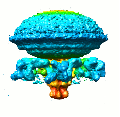
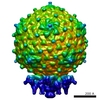
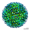
| Title | The structures of bacteriophages K1E and K1-5 explain processive degradation of polysaccharide capsules and evolution of new host specificities. | |||||||||
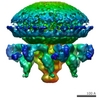
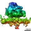
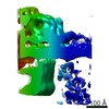
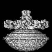
 Map data Map data | Sixfold averaged tail of bacteriophage K1E | |||||||||
 Sample Sample |
| |||||||||
| Biological species |  Enterobacteria phage K1E (virus) Enterobacteria phage K1E (virus) | |||||||||
| Method |  single particle reconstruction / cryo EM / negative staining / Resolution: 16.4 Å single particle reconstruction / cryo EM / negative staining / Resolution: 16.4 Å | |||||||||
 Authors Authors | Leiman PG / Battisti AJ / Bowman VD / Stummeyer K / Muhlenhoff M / Gerardy-Schahn R / Scholl D / Molineux IJ | |||||||||
 Citation Citation | Journal: J Mol Biol / Year: 2007 Title: The structures of bacteriophages K1E and K1-5 explain processive degradation of polysaccharide capsules and evolution of new host specificities. Authors: Petr G Leiman / Anthony J Battisti / Valorie D Bowman / Katharina Stummeyer / Martina Mühlenhoff / Rita Gerardy-Schahn / Dean Scholl / Ian J Molineux /  Abstract: External polysaccharides of many pathogenic bacteria form capsules protecting the bacteria from the animal immune system and phage infection. However, some bacteriophages can digest these capsules ...External polysaccharides of many pathogenic bacteria form capsules protecting the bacteria from the animal immune system and phage infection. However, some bacteriophages can digest these capsules using glycosidases displayed on the phage particle. We have utilized cryo-electron microscopy to determine the structures of phages K1E and K1-5 and thereby establish the mechanism by which these phages attain and switch their host specificity. Using a specific glycosidase, both phages penetrate the capsule and infect the neuroinvasive human pathogen Escherichia coli K1. In addition to the K1-specific glycosidase, each K1-5 particle carries a second enzyme that allows it to infect E. coli K5, whose capsule is chemically different from that of K1. The enzymes are organized into a multiprotein complex attached via an adapter protein to the virus portal vertex, through which the DNA is ejected during infection. The structure of the complex suggests a mechanism for the apparent processivity of degradation that occurs as the phage drills through the polysaccharide capsule. The enzymes recognize the adapter protein by a conserved N-terminal sequence, providing a mechanism for phages to acquire different enzymes and thus to evolve new host specificities. | |||||||||
| History |
|
- Structure visualization
| Movie |
 Movie viewer Movie viewer |
|---|---|
| Structure viewer | EM map: SurfViewMolmilJmol/JSmol |
| Supplemental images |
- Downloads & links
-EMDB archive
| Map data | emd_1333.map.gz | 13.1 MB | EMDB map data format | |
|---|---|---|---|---|
| Header (meta data) | emd-1333-v30.xmlemd-1333.xml | 9.4 KB 9.4 KB | Display Display | EMDB header |
| Images |  1333.gif 1333.gif | 51.3 KB | ||
| Archive directory |  http://ftp.pdbj.org/pub/emdb/structures/EMD-1333ftp://ftp.pdbj.org/pub/emdb/structures/EMD-1333 http://ftp.pdbj.org/pub/emdb/structures/EMD-1333ftp://ftp.pdbj.org/pub/emdb/structures/EMD-1333 | HTTPS FTP |
-Related structure data
-Links
| EMDB pages | EMDB (EBI/PDBe) / EMDataResource |
|---|
-Map
| File | Download / File: emd_1333.map.gz / Format: CCP4 / Size: 20.3 MB / Type: IMAGE STORED AS FLOATING POINT NUMBER (4 BYTES) | ||||||||||||||||||||||||||||||||||||||||||||||||||||||||||||||||||||
|---|---|---|---|---|---|---|---|---|---|---|---|---|---|---|---|---|---|---|---|---|---|---|---|---|---|---|---|---|---|---|---|---|---|---|---|---|---|---|---|---|---|---|---|---|---|---|---|---|---|---|---|---|---|---|---|---|---|---|---|---|---|---|---|---|---|---|---|---|---|
| Annotation | Sixfold averaged tail of bacteriophage K1E | ||||||||||||||||||||||||||||||||||||||||||||||||||||||||||||||||||||
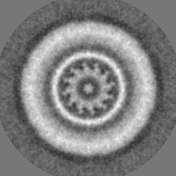
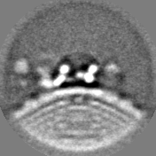
| Projections & slices | Image control
Images are generated by Spider. | ||||||||||||||||||||||||||||||||||||||||||||||||||||||||||||||||||||
| Voxel size | X=Y=Z: 2.98 Å | ||||||||||||||||||||||||||||||||||||||||||||||||||||||||||||||||||||
| Density |
| ||||||||||||||||||||||||||||||||||||||||||||||||||||||||||||||||||||
| Symmetry | Space group: 1 | ||||||||||||||||||||||||||||||||||||||||||||||||||||||||||||||||||||
| Details | EMDB XML:
CCP4 map header:
| ||||||||||||||||||||||||||||||||||||||||||||||||||||||||||||||||||||
 Z (Sec.)
 Y (Row.)
 X (Col.)


-Supplemental data
- Sample components
-Entire : Sixfold averaged tail of bacteriophage K1E
| Entire | Name: Sixfold averaged tail of bacteriophage K1E |
|---|---|
| Components |
|
-Supramolecule #1000: Sixfold averaged tail of bacteriophage K1E
| Supramolecule | Name: Sixfold averaged tail of bacteriophage K1E / type: sample / ID: 1000 / Number unique components: 1 |
|---|---|
| Molecular weight | Experimental: 8 MDa / Theoretical: 8 MDa / Method: Primary sequence |
-Supramolecule #1: Enterobacteria phage K1E
| Supramolecule | Name: Enterobacteria phage K1E / type: virus / ID: 1 / Name.synonym: phage K1E tail / Details: sixfold averaged / NCBI-ID: 344022 / Sci species name: Enterobacteria phage K1E / Virus type: VIRION / Virus isolate: SPECIES / Virus enveloped: No / Virus empty: No / Syn species name: phage K1E tail |
|---|---|
| Host (natural) | Organism:  Escherichia coli (E. coli) / synonym: BACTERIA(EUBACTERIA) Escherichia coli (E. coli) / synonym: BACTERIA(EUBACTERIA) |
| Molecular weight | Experimental: 8 MDa / Theoretical: 8 MDa |
-Experimental details
-Structure determination
| Method | negative staining, cryo EM |
|---|---|
 Processing Processing | single particle reconstruction |
| Aggregation state | particle |
-Sample preparation
| Concentration | 1 mg/mL |
|---|---|
| Buffer | pH: 7.5 / Details: 50 mM Tris HCl pH 7.5, 100 mM NaCl, 8 mM MgSO4 |
| Staining | Type: NEGATIVE / Details: none |
| Grid | Details: quantifoil grid |
| Vitrification | Cryogen name: ETHANE / Chamber humidity: 80 % / Chamber temperature: 300 K / Instrument: OTHER / Details: Vitrification instrument: Vitrobot |
- Electron microscopy
| Microscope | FEI/PHILIPS CM300FEG/T |
|---|---|
| Electron beam | Acceleration voltage: 300 kV / Electron source: FIELD EMISSION GUN |
| Electron optics | Calibrated magnification: 47000 / Illumination mode: SPOT SCAN / Imaging mode: BRIGHT FIELDBright-field microscopy / Cs: 2.0 mm / Nominal defocus max: 3.3 µm / Nominal defocus min: 0.7 µm / Nominal magnification: 45000 |
| Sample stage | Specimen holder: Side entry liquid nitrogen-cooled cryo specimen holder Specimen holder model: GATAN LIQUID NITROGEN |
| Temperature | Average: 100 K |
| Date | May 10, 2005 |
| Image recording | Category: FILM / Film or detector model: KODAK SO-163 FILM / Digitization - Scanner: ZEISS SCAI / Digitization - Sampling interval: 7 µm / Number real images: 122 / Average electron dose: 25 e/Å2 / Bits/pixel: 12 |
-Image processing
| CTF correction | Details: Each particle |
|---|---|
| Final reconstruction | Applied symmetry - Point group: C6 (6 fold cyclic) / Algorithm: OTHER / Resolution.type: BY AUTHOR / Resolution: 16.4 Å / Resolution method: OTHER / Software - Name: Spider / Number images used: 6105 |
