 Movie
Movie Controller


+ Open data

- Basic information
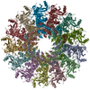
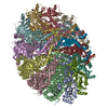
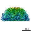
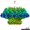
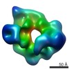
| Entry | Database: PDB / ID: 5jzh | ||||||
|---|---|---|---|---|---|---|---|
| Title | Cryo-EM structure of aerolysin prepore | ||||||
 Components Components | Aerolysin | ||||||
 Keywords Keywords | TOXIN / pore forming toxin / concentric beta-barrel / aerolysin | ||||||
| Function / homology |  Function and homology information Function and homology informationtoxin activity / host cell plasma membrane / extracellular region / identical protein binding / membrane Similarity search - Function | ||||||
| Biological species |  Aeromonas hydrophila (bacteria) Aeromonas hydrophila (bacteria) | ||||||
| Method | ELECTRON MICROSCOPY / single particle reconstruction / cryo EM / Resolution: 3.9 Å | ||||||
 Authors Authors | Iacovache, I. / Zuber, B. | ||||||
| Funding support |  Switzerland, 1items Switzerland, 1items
| ||||||
 Citation Citation | Journal: Nat Commun / Year: 2016 Title: Cryo-EM structure of aerolysin variants reveals a novel protein fold and the pore-formation process. Authors: Ioan Iacovache / Sacha De Carlo / Nuria Cirauqui / Matteo Dal Peraro / F Gisou van der Goot / Benoît Zuber /   Abstract: Owing to their pathogenical role and unique ability to exist both as soluble proteins and transmembrane complexes, pore-forming toxins (PFTs) have been a focus of microbiologists and structural ...Owing to their pathogenical role and unique ability to exist both as soluble proteins and transmembrane complexes, pore-forming toxins (PFTs) have been a focus of microbiologists and structural biologists for decades. PFTs are generally secreted as water-soluble monomers and subsequently bind the membrane of target cells. Then, they assemble into circular oligomers, which undergo conformational changes that allow membrane insertion leading to pore formation and potentially cell death. Aerolysin, produced by the human pathogen Aeromonas hydrophila, is the founding member of a major PFT family found throughout all kingdoms of life. We report cryo-electron microscopy structures of three conformational intermediates and of the final aerolysin pore, jointly providing insight into the conformational changes that allow pore formation. Moreover, the structures reveal a protein fold consisting of two concentric β-barrels, tightly kept together by hydrophobic interactions. This fold suggests a basis for the prion-like ultrastability of aerolysin pore and its stoichiometry. | ||||||
| History |
|
- Structure visualization
| Movie |
Movie viewer |
|---|---|
| Structure viewer | Molecule: MolmilJmol/JSmol |
- Downloads & links
-Download
| PDBx/mmCIF format | 5jzh.cif.gz | 1.8 MB | Display | PDBx/mmCIF format |
|---|---|---|---|---|
| PDB format | pdb5jzh.ent.gz | 1.5 MB | Display | PDB format |
| PDBx/mmJSON format | 5jzh.json.gz | Tree view | PDBx/mmJSON format | |
| Others |  Other downloads Other downloads |
-Validation report
| Summary document | 5jzh_validation.pdf.gz | 893.2 KB | Display | wwPDB validaton report |
|---|---|---|---|---|
| Full document | 5jzh_full_validation.pdf.gz | 941.8 KB | Display | |
| Data in XML | 5jzh_validation.xml.gz | 148.9 KB | Display | |
| Data in CIF | 5jzh_validation.cif.gz | 223.6 KB | Display | |
| Arichive directory | https://data.pdbj.org/pub/pdb/validation_reports/jz/5jzhftp://data.pdbj.org/pub/pdb/validation_reports/jz/5jzh | HTTPS FTP |
-Related structure data
| Related structure data | 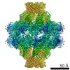 8185MC 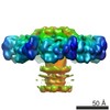 8187C 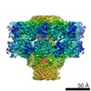 8188C  5jztC  5jzwC M: map data used to model this data C: citing same article ( |
|---|---|
| Similar structure data |

-Links
 PDBj
PDBj


- Assembly
| Deposited unit | 
|
|---|---|
| 1 | 
|
| 2 | 
|
-Components
| #1: Protein | Mass: 47079.031 Da / Num. of mol.: 14 / Mutation: Y221G Source method: isolated from a genetically manipulated source Source: (gene. exp.) Aeromonas hydrophila (bacteria) / Gene: aerA / Production host: |
|---|
-Experimental details
-Experiment
| Experiment | Method: ELECTRON MICROSCOPY |
|---|---|
| EM experiment | Aggregation state: PARTICLE / 3D reconstruction method: single particle reconstruction |
- Sample preparation
| Component | Name: aerolysin prepore / Type: COMPLEX Details: Aerolysin oligomer arrested in it's prepore conformation by mutagenesis (Y221G). Entity ID: all / Source: RECOMBINANT |
|---|---|
| Molecular weight | Value: 0.35 MDa / Experimental value: NO |
| Source (natural) | Organism: Aeromonas hydrophila (bacteria) |
| Source (recombinant) | Organism: |
| Buffer solution | pH: 7.4 |
| Specimen | Conc.: 0.4 mg/ml / Embedding applied: NO / Shadowing applied: NO / Staining applied: NO / Vitrification applied: YES |
| Specimen support | Grid material: COPPER / Grid mesh size: 300 divisions/in. / Grid type: Quantifoil R2/1 |
| Vitrification | Instrument: FEI VITROBOT MARK IV / Cryogen name: ETHANE / Humidity: 70 % / Chamber temperature: 295 K Details: 30 seconds glow discharged grids, 4ul sample, 5 seconds blot |
- Electron microscopy imaging
| Experimental equipment |  Model: Titan Krios / Image courtesy: FEI Company |
|---|---|
| Microscopy | Model: FEI TITAN KRIOS |
| Electron gun | Electron source:  FIELD EMISSION GUN / Accelerating voltage: 300 kV / Illumination mode: FLOOD BEAM FIELD EMISSION GUN / Accelerating voltage: 300 kV / Illumination mode: FLOOD BEAM |
| Electron lens | Mode: BRIGHT FIELD |
| Image recording | Average exposure time: 1 sec. / Electron dose: 22.42 e/Å2 / Detector mode: INTEGRATING / Film or detector model: FEI FALCON II (4k x 4k) |
- Processing
| EM software |
| ||||||||||||||||||||||||||||||||||||||||
|---|---|---|---|---|---|---|---|---|---|---|---|---|---|---|---|---|---|---|---|---|---|---|---|---|---|---|---|---|---|---|---|---|---|---|---|---|---|---|---|---|---|
| CTF correction | Type: PHASE FLIPPING AND AMPLITUDE CORRECTION | ||||||||||||||||||||||||||||||||||||||||
| Particle selection | Num. of particles selected: 56966 | ||||||||||||||||||||||||||||||||||||||||
| Symmetry | Point symmetry: D7 (2x7 fold dihedral) | ||||||||||||||||||||||||||||||||||||||||
| 3D reconstruction | Resolution: 3.9 Å / Resolution method: FSC 0.143 CUT-OFF / Num. of particles: 42962 / Symmetry type: POINT | ||||||||||||||||||||||||||||||||||||||||
| Atomic model building | Protocol: FLEXIBLE FIT |
