Movie
Movie Controller


+ Open data

- Basic information
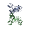
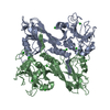
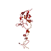
| Entry | Database: PDB / ID: 5hx2 | ||||||
|---|---|---|---|---|---|---|---|
| Title | In vitro assembled star-shaped hubless T4 baseplate | ||||||
 Components Components |
| ||||||
 Keywords Keywords | VIRAL PROTEIN / T4 / baseplate / complex | ||||||
| Function / homology |  Function and homology information Function and homology informationvirus tail, baseplate / viral tail assembly / viral release from host cell / identical protein binding Similarity search - Function | ||||||
| Biological species |  Enterobacteria phage T4 (virus) Enterobacteria phage T4 (virus) | ||||||
| Method | ELECTRON MICROSCOPY / single particle reconstruction / cryo EM / Resolution: 3.8 Å | ||||||
 Authors Authors | Yap, M.L. / Klose, T. / Fokine, A. / Rossmann, M.G. | ||||||
| Funding support |  United States, 1items United States, 1items
| ||||||
 Citation Citation | Journal: Proc Natl Acad Sci U S A / Year: 2016 Title: Role of bacteriophage T4 baseplate in regulating assembly and infection. Authors: Moh Lan Yap / Thomas Klose / Fumio Arisaka / Jeffrey A Speir / David Veesler / Andrei Fokine / Michael G Rossmann /  Abstract: Bacteriophage T4 consists of a head for protecting its genome and a sheathed tail for inserting its genome into a host. The tail terminates with a multiprotein baseplate that changes its conformation ...Bacteriophage T4 consists of a head for protecting its genome and a sheathed tail for inserting its genome into a host. The tail terminates with a multiprotein baseplate that changes its conformation from a "high-energy" dome-shaped to a "low-energy" star-shaped structure during infection. Although these two structures represent different minima in the total energy landscape of the baseplate assembly, as the dome-shaped structure readily changes to the star-shaped structure when the virus infects a host bacterium, the dome-shaped structure must have more energy than the star-shaped structure. Here we describe the electron microscopy structure of a 3.3-MDa in vitro-assembled star-shaped baseplate with a resolution of 3.8 Å. This structure, together with other genetic and structural data, shows why the high-energy baseplate is formed in the presence of the central hub and how the baseplate changes to the low-energy structure, via two steps during infection. Thus, the presence of the central hub is required to initiate the assembly of metastable, high-energy structures. If the high-energy structure is formed and stabilized faster than the low-energy structure, there will be insufficient components to assemble the low-energy structure. | ||||||
| History |
|
- Structure visualization
| Movie |
Movie viewer |
|---|---|
| Structure viewer | Molecule: MolmilJmol/JSmol |
- Downloads & links
-Download
| PDBx/mmCIF format | 5hx2.cif.gz | 750.7 KB | Display | PDBx/mmCIF format |
|---|---|---|---|---|
| PDB format | pdb5hx2.ent.gz | 572.1 KB | Display | PDB format |
| PDBx/mmJSON format | 5hx2.json.gz | Tree view | PDBx/mmJSON format | |
| Others |  Other downloads Other downloads |
-Validation report
| Summary document | 5hx2_validation.pdf.gz | 852.6 KB | Display | wwPDB validaton report |
|---|---|---|---|---|
| Full document | 5hx2_full_validation.pdf.gz | 998.2 KB | Display | |
| Data in XML | 5hx2_validation.xml.gz | 134 KB | Display | |
| Data in CIF | 5hx2_validation.cif.gz | 200.4 KB | Display | |
| Arichive directory | https://data.pdbj.org/pub/pdb/validation_reports/hx/5hx2ftp://data.pdbj.org/pub/pdb/validation_reports/hx/5hx2 | HTTPS FTP |
-Related structure data
| Related structure data | 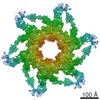 8064MC M: map data used to model this data C: citing same article ( |
|---|---|
| Similar structure data |
-Links
 PDBj
PDBj- Assembly
| Deposited unit | 
|
|---|---|
| 1 | x 6
|
| 2 |
|
| 3 | 
|
| Symmetry | Point symmetry: (Schoenflies symbol: C6 (6 fold cyclic)) |
-Components
| #1: Protein | Mass: 119336.516 Da / Num. of mol.: 1 Source method: isolated from a genetically manipulated source Source: (gene. exp.) Enterobacteria phage T4 (virus) / Gene: 7 / Plasmid: pET29 / Production host:  | ||||||
|---|---|---|---|---|---|---|---|
| #2: Protein | Mass: 38041.668 Da / Num. of mol.: 2 Source method: isolated from a genetically manipulated source Source: (gene. exp.) Enterobacteria phage T4 (virus) / Gene: 8 / Plasmid: pET29 / Production host: #3: Protein | Mass: 74492.641 Da / Num. of mol.: 2 Source method: isolated from a genetically manipulated source Source: (gene. exp.) Enterobacteria phage T4 (virus) / Gene: 6 / Plasmid: pET29 / Production host: #4: Protein | | Mass: 22990.885 Da / Num. of mol.: 1 Source method: isolated from a genetically manipulated source Source: (gene. exp.) Enterobacteria phage T4 (virus) / Gene: 53 / Plasmid: pET29 / Production host: #5: Protein | Mass: 66281.680 Da / Num. of mol.: 3 Source method: isolated from a genetically manipulated source Source: (gene. exp.) Enterobacteria phage T4 (virus) / Gene: 10 / Plasmid: pET29 / Production host: |
-Experimental details
-Experiment
| Experiment | Method: ELECTRON MICROSCOPY |
|---|---|
| EM experiment | Aggregation state: PARTICLE / 3D reconstruction method: single particle reconstruction |
- Sample preparation
| Component | Name: In vitro assembled hubless T4 baseplate / Type: COMPLEX / Entity ID: all / Source: RECOMBINANT |
|---|---|
| Molecular weight | Value: 3.3 MDa / Experimental value: NO |
| Source (natural) | Organism: Enterobacteria phage T4 (virus) |
| Source (recombinant) | Organism: |
| Buffer solution | pH: 8 |
| Specimen | Conc.: 2 mg/ml / Embedding applied: NO / Shadowing applied: NO / Staining applied: NO / Vitrification applied: YES |
| Specimen support | Details: 400-mesh copper CF-1.2/1.3-4C / Grid material: COPPER / Grid mesh size: 400 divisions/in. / Grid type: CF-1.2/1.3-4C |
| Vitrification | Instrument: GATAN CRYOPLUNGE 3 / Cryogen name: ETHANE / Humidity: 80 % / Details: Plunged into liquid ethane (GATAN CRYOPLUNGE 3). |
- Electron microscopy imaging
| Experimental equipment |  Model: Titan Krios / Image courtesy: FEI Company |
|---|---|
| Microscopy | Model: FEI TITAN KRIOS |
| Electron gun | Electron source:  FIELD EMISSION GUN / Accelerating voltage: 300 kV / Illumination mode: OTHER FIELD EMISSION GUN / Accelerating voltage: 300 kV / Illumination mode: OTHER |
| Electron lens | Mode: BRIGHT FIELD / Nominal magnification: 22500 X / Calibrated magnification: 38168 X / Nominal defocus max: 3500 nm / Nominal defocus min: 500 nm / Calibrated defocus min: 400 nm / Calibrated defocus max: 3740 nm / Cs: 2.7 mm / Alignment procedure: ZEMLIN TABLEAU |
| Specimen holder | Cryogen: NITROGEN / Specimen holder model: FEI TITAN KRIOS AUTOGRID HOLDER |
| Image recording | Average exposure time: 7.6 sec. / Electron dose: 35 e/Å2 / Detector mode: SUPER-RESOLUTION / Film or detector model: GATAN K2 SUMMIT (4k x 4k) / Num. of grids imaged: 2 / Num. of real images: 1725 |
| Image scans | Width: 7676 / Height: 7420 / Movie frames/image: 38 / Used frames/image: 3-38 |
- Processing
| EM software |
| ||||||||||||||||||||||||||||||||||||||||||||||||||
|---|---|---|---|---|---|---|---|---|---|---|---|---|---|---|---|---|---|---|---|---|---|---|---|---|---|---|---|---|---|---|---|---|---|---|---|---|---|---|---|---|---|---|---|---|---|---|---|---|---|---|---|
| CTF correction | Type: PHASE FLIPPING AND AMPLITUDE CORRECTION | ||||||||||||||||||||||||||||||||||||||||||||||||||
| Particle selection | Num. of particles selected: 69920 | ||||||||||||||||||||||||||||||||||||||||||||||||||
| Symmetry | Point symmetry: C6 (6 fold cyclic) | ||||||||||||||||||||||||||||||||||||||||||||||||||
| 3D reconstruction | Resolution: 3.8 Å / Resolution method: FSC 0.143 CUT-OFF / Num. of particles: 45607 / Symmetry type: POINT | ||||||||||||||||||||||||||||||||||||||||||||||||||
| Atomic model building | Protocol: AB INITIO MODEL / Space: REAL | ||||||||||||||||||||||||||||||||||||||||||||||||||
| Atomic model building |
|