 Movie
Movie Controller


+ Open data

- Basic information
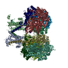
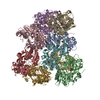
| Entry | Database: PDB / ID: 5gao | ||||||
|---|---|---|---|---|---|---|---|
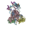
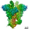
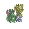
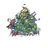
| Title | Head region of the yeast spliceosomal U4/U6.U5 tri-snRNP | ||||||
 Components Components |
| ||||||
 Keywords Keywords |  TRANSCRIPTION / pre-mRNA splicing / snRNP / GTPase / spliceosome TRANSCRIPTION / pre-mRNA splicing / snRNP / GTPase / spliceosome | ||||||
| Function / homology |  Function and homology information Function and homology informationmaturation of 5S rRNA / spliceosome conformational change to release U4 (or U4atac) and U1 (or U11) / splicing factor binding / U4/U6 snRNP / 7-methylguanosine cap hypermethylation / pICln-Sm protein complex / spliceosomal tri-snRNP complex / small nuclear ribonucleoprotein complex / SMN-Sm protein complex / mRNA cis splicing, via spliceosome ...maturation of 5S rRNA / spliceosome conformational change to release U4 (or U4atac) and U1 (or U11) / splicing factor binding / U4/U6 snRNP / 7-methylguanosine cap hypermethylation / pICln-Sm protein complex / spliceosomal tri-snRNP complex / small nuclear ribonucleoprotein complex / SMN-Sm protein complex / mRNA cis splicing, via spliceosome / U2-type prespliceosome assembly / commitment complex / U4 snRNP / U2 snRNP / poly(U) RNA binding / U1 snRNP / U2-type prespliceosome / precatalytic spliceosome / spliceosomal complex assembly / generation of catalytic spliceosome for second transesterification step / mRNA 3'-splice site recognition / mRNA 5'-splice site recognition / spliceosomal tri-snRNP complex assembly / U5 snRNA binding / U5 snRNP / U2 snRNA binding / spliceosomal snRNP assembly / U6 snRNA binding / pre-mRNA intronic binding / U1 snRNA binding / U4/U6 x U5 tri-snRNP complex / catalytic step 2 spliceosome / spliceosomal complex / mRNA splicing, via spliceosome / metallopeptidase activity / RNA helicase activity / nucleic acid binding / RNA helicase / mRNA binding / ATP hydrolysis activity / RNA binding / ATP binding / identical protein binding / nucleus / cytosol / cytoplasmSimilarity search - Function | ||||||
| Biological species |  Saccharomyces cerevisiae (brewer's yeast) Saccharomyces cerevisiae (brewer's yeast) | ||||||
| Method | ELECTRON MICROSCOPY / single particle reconstruction / Resolution: 4.2 Å | ||||||
 Authors Authors | Nguyen, T.H.D. / Galej, W.P. / Bai, X.C. / Oubridge, C. / Scheres, S.H.W. / Newman, A.J. / Nagai, K. | ||||||
| Funding support |  United Kingdom, 1items United Kingdom, 1items
| ||||||
 Citation Citation | Journal: Nature / Year: 2016 Title: Cryo-EM structure of the yeast U4/U6.U5 tri-snRNP at 3.7 Å resolution. Authors: Thi Hoang Duong Nguyen / Wojciech P Galej / Xiao-Chen Bai / Chris Oubridge / Andrew J Newman / Sjors H W Scheres / Kiyoshi Nagai / Abstract: U4/U6.U5 tri-snRNP represents a substantial part of the spliceosome before activation. A cryo-electron microscopy structure of Saccharomyces cerevisiae U4/U6.U5 tri-snRNP at 3.7 Å resolution led ...U4/U6.U5 tri-snRNP represents a substantial part of the spliceosome before activation. A cryo-electron microscopy structure of Saccharomyces cerevisiae U4/U6.U5 tri-snRNP at 3.7 Å resolution led to an essentially complete atomic model comprising 30 proteins plus U4/U6 and U5 small nuclear RNAs (snRNAs). The structure reveals striking interweaving interactions of the protein and RNA components, including extended polypeptides penetrating into subunit interfaces. The invariant ACAGAGA sequence of U6 snRNA, which base-pairs with the 5'-splice site during catalytic activation, forms a hairpin stabilized by Dib1 and Prp8 while the adjacent nucleotides interact with the exon binding loop 1 of U5 snRNA. Snu114 harbours GTP, but its putative catalytic histidine is held away from the γ-phosphate by hydrogen bonding to a tyrosine in the amino-terminal domain of Prp8. Mutation of this histidine to alanine has no detectable effect on yeast growth. The structure provides important new insights into the spliceosome activation process leading to the formation of the catalytic centre. #1: Journal: Nature / Year: 2015Title: The architecture of the spliceosomal U4/U6.U5 tri-snRNP. Authors: Thi Hoang Duong Nguyen / Wojciech P Galej / Xiao-chen Bai / Christos G Savva / Andrew J Newman / Sjors H W Scheres / Kiyoshi Nagai / Abstract: U4/U6.U5 tri-snRNP is a 1.5-megadalton pre-assembled spliceosomal complex comprising U5 small nuclear RNA (snRNA), extensively base-paired U4/U6 snRNAs and more than 30 proteins, including the key ...U4/U6.U5 tri-snRNP is a 1.5-megadalton pre-assembled spliceosomal complex comprising U5 small nuclear RNA (snRNA), extensively base-paired U4/U6 snRNAs and more than 30 proteins, including the key components Prp8, Brr2 and Snu114. The tri-snRNP combines with a precursor messenger RNA substrate bound to U1 and U2 small nuclear ribonucleoprotein particles (snRNPs), and transforms into a catalytically active spliceosome after extensive compositional and conformational changes triggered by unwinding of the U4 and U6 (U4/U6) snRNAs. Here we use cryo-electron microscopy single-particle reconstruction of Saccharomyces cerevisiae tri-snRNP at 5.9 Å resolution to reveal the essentially complete organization of its RNA and protein components. The single-stranded region of U4 snRNA between its 3' stem-loop and the U4/U6 snRNA stem I is loaded into the Brr2 helicase active site ready for unwinding. Snu114 and the amino-terminal domain of Prp8 position U5 snRNA to insert its loop I, which aligns the exons for splicing, into the Prp8 active site cavity. The structure provides crucial insights into the activation process and the active site of the spliceosome. | ||||||
| History |
|
- Structure visualization
| Movie |
Movie viewer |
|---|---|
| Structure viewer | Molecule: MolmilJmol/JSmol |
- Downloads & links
-Download
| PDBx/mmCIF format | 5gao.cif.gz | 592.9 KB | Display | PDBx/mmCIF format |
|---|---|---|---|---|
| PDB format | pdb5gao.ent.gz | 475.2 KB | Display | PDB format |
| PDBx/mmJSON format | 5gao.json.gz | Tree view | PDBx/mmJSON format | |
| Others |  Other downloads Other downloads |
-Validation report
| Arichive directory | https://data.pdbj.org/pub/pdb/validation_reports/ga/5gaoftp://data.pdbj.org/pub/pdb/validation_reports/ga/5gao | HTTPS FTP |
|---|
-Related structure data
| Related structure data | 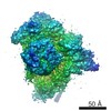 8013MC 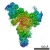 8006C 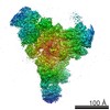 8007C 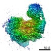 8008C  8009C  8010C 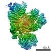 8011C 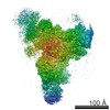 8012C 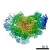 8014C 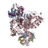 5gamC 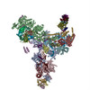 5ganC 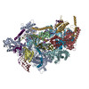 5gapC M: map data used to model this data C: citing same article ( |
|---|---|
| Similar structure data |

-Links
 PDBj
PDBj


- Assembly
| Deposited unit | 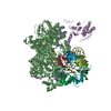
|
|---|---|
| 1 |
|
-Components
-Protein , 2 types, 2 molecules kE
| #1: Protein | Mass: 22426.990 Da / Num. of mol.: 1 / Source method: isolated from a natural source Details: The SmB protein from the U4 snRNP Sm protein ring. The C-terminus is disordered in this and other previously reported structures. Source: (natural) Saccharomyces cerevisiae (brewer's yeast) / Strain: BCY123 / References: UniProt: P40018 |
|---|---|
| #8: Protein | Mass: 25078.721 Da / Num. of mol.: 1 / Source method: isolated from a natural source Details: NB: Snu66 protein has been mostly fitted as poly(Ala) into helices and extended polypeptide within the EM map. The authors do not believe the numbering to be accurate and it is likely incorrect. Source: (natural) Saccharomyces cerevisiae (brewer's yeast) / Strain: BCY123 / References: UniProt: Q12420*PLUS |
-Small nuclear ribonucleoprotein ... , 6 types, 6 molecules lmnpqr
| #2: Protein | Mass: 16296.798 Da / Num. of mol.: 1 / Source method: isolated from a natural source Details: The SmD1 protein from the U4 snRNP Sm protein ring. Source: (natural) Saccharomyces cerevisiae (brewer's yeast) / Strain: BCY123 / References: UniProt: Q02260 |
|---|---|
| #3: Protein | Mass: 12876.066 Da / Num. of mol.: 1 / Source method: isolated from a natural source Details: The SmD2 protein from the U4 snRNP Sm protein ring. Source: (natural) Saccharomyces cerevisiae (brewer's yeast) / Strain: BCY123 / References: UniProt: Q06217 |
| #4: Protein | Mass: 11240.139 Da / Num. of mol.: 1 / Source method: isolated from a natural source Details: The SmD3 protein from the U4 snRNP Sm protein ring. Source: (natural) Saccharomyces cerevisiae (brewer's yeast) / Strain: BCY123 / References: UniProt: P43321 |
| #5: Protein | Mass: 10385.098 Da / Num. of mol.: 1 / Source method: isolated from a natural source / Details: The SmE protein from the U4 snRNP Sm protein ring. / Source: (natural) Saccharomyces cerevisiae (brewer's yeast) / Strain: BCY123 / References: UniProt: Q12330 |
| #6: Protein | Mass: 9669.945 Da / Num. of mol.: 1 / Source method: isolated from a natural source / Details: The SmF protein from the U4 snRNP Sm protein ring. / Source: (natural) Saccharomyces cerevisiae (brewer's yeast) / Strain: BCY123 / References: UniProt: P54999 |
| #7: Protein | Mass: 8490.809 Da / Num. of mol.: 1 / Source method: isolated from a natural source / Details: The SmG protein from the U4 snRNP Sm protein ring. / Source: (natural) Saccharomyces cerevisiae (brewer's yeast) / Strain: BCY123 / References: UniProt: P40204 |
-Pre-mRNA-splicing ... , 2 types, 2 molecules BA
| #9: Protein | Mass: 246470.266 Da / Num. of mol.: 1 / Source method: isolated from a natural source Details: Brr2 RNA helicase loaded onto U4 snRNA strand of the U4/U6 duplex between Stem 1 duplex and the 3' stem loop. Source: (natural) Saccharomyces cerevisiae (brewer's yeast) / Strain: BCY123 / References: UniProt: P32639, RNA helicase |
|---|---|
| #11: Protein | Mass: 30231.008 Da / Num. of mol.: 1 / Source method: isolated from a natural source Details: Prp8 Jab1/MPN domain is connected to the rest of the Prp8 protein by a flexible linker peptide. Source: (natural) Saccharomyces cerevisiae (brewer's yeast) / Strain: BCY123 / References: UniProt: P33334 |
-RNA chain , 1 types, 1 molecules V
| #10: RNA chain | Mass: 30615.994 Da / Num. of mol.: 1 / Source method: isolated from a natural source Details: This U4 snRNA region has a disordered region between stem 1 of the U4/U6 snRNA duplex and where the RNA is loaded into Brr2 protein. The apical part of the 3' stem loop and the region ...Details: This U4 snRNA region has a disordered region between stem 1 of the U4/U6 snRNA duplex and where the RNA is loaded into Brr2 protein. The apical part of the 3' stem loop and the region following the Sm site are also disordered. Source: (natural) Saccharomyces cerevisiae (brewer's yeast) / Strain: BCY123 / References: GenBank: 807071957 |
|---|
-Experimental details
-Experiment
| Experiment | Method: ELECTRON MICROSCOPY |
|---|---|
| EM experiment | Aggregation state: PARTICLE / 3D reconstruction method: single particle reconstruction |
- Sample preparation
| Component | Name: Head region of the yeast spliceosomal U4/U6.U5 tri-snRNP Type: COMPLEX / Entity ID: all / Source: NATURAL |
|---|---|
| Molecular weight | Value: 0.40 MDa / Experimental value: NO |
| Source (natural) | Organism: Saccharomyces cerevisiae (brewer's yeast) / Strain: BCY123 |
| Buffer solution | pH: 7.9 |
| Buffer component | Conc.: 1 mM / Name: DTT |
| Specimen | Conc.: 0.2 mg/ml / Embedding applied: NO / Shadowing applied: NO / Staining applied: NO / Vitrification applied: NO |
| Specimen support | Details: Grids are made of holey carbon, carbon-coated and glow discharged in N-amylamine. Grid material: COPPER / Grid mesh size: 400 divisions/in. / Grid type: Quantifoil R1.2/1.3 |
- Electron microscopy imaging
| Experimental equipment |  Model: Titan Krios / Image courtesy: FEI Company |
|---|---|
| Microscopy | Model: FEI TITAN KRIOS |
| Electron gun | Electron source: FIELD EMISSION GUN / Accelerating voltage: 300 kV / Illumination mode: FLOOD BEAM |
| Electron lens | Mode: BRIGHT FIELDBright-field microscopy / Nominal magnification: 81000 X / Calibrated magnification: 35714 X / Nominal defocus max: 3500 nm / Nominal defocus min: 500 nm / Cs: 2 mm |
| Specimen holder | Cryogen: NITROGEN / Specimen holder model: FEI TITAN KRIOS AUTOGRID HOLDER |
| Image recording | Average exposure time: 16 sec. / Electron dose: 38 e/Å2 / Detector mode: SUPER-RESOLUTION / Film or detector model: GATAN K2 SUMMIT (4k x 4k) / Num. of real images: 2477 |
| EM imaging optics | Energyfilter name: GIF Quantum |
| Image scans | Movie frames/image: 20 / Used frames/image: 1-20 |
- Processing
| Software | Name: REFMAC / Version: 5.8.0124 / Classification: refinement | ||||||||||||||||||||||||||||||||||||||||||||||||||||||||||||||||||||||||||||||||||||||||||||||||||||||||||
|---|---|---|---|---|---|---|---|---|---|---|---|---|---|---|---|---|---|---|---|---|---|---|---|---|---|---|---|---|---|---|---|---|---|---|---|---|---|---|---|---|---|---|---|---|---|---|---|---|---|---|---|---|---|---|---|---|---|---|---|---|---|---|---|---|---|---|---|---|---|---|---|---|---|---|---|---|---|---|---|---|---|---|---|---|---|---|---|---|---|---|---|---|---|---|---|---|---|---|---|---|---|---|---|---|---|---|---|
| EM software |
| ||||||||||||||||||||||||||||||||||||||||||||||||||||||||||||||||||||||||||||||||||||||||||||||||||||||||||
| CTF correction | Type: PHASE FLIPPING ONLY | ||||||||||||||||||||||||||||||||||||||||||||||||||||||||||||||||||||||||||||||||||||||||||||||||||||||||||
| Particle selection | Num. of particles selected: 473827 | ||||||||||||||||||||||||||||||||||||||||||||||||||||||||||||||||||||||||||||||||||||||||||||||||||||||||||
| 3D reconstruction | Resolution: 4.2 Å / Resolution method: FSC 0.143 CUT-OFF / Num. of particles: 140155 / Num. of class averages: 1 / Symmetry type: POINT | ||||||||||||||||||||||||||||||||||||||||||||||||||||||||||||||||||||||||||||||||||||||||||||||||||||||||||
| Atomic model building | Protocol: OTHER / Space: RECIPROCAL | ||||||||||||||||||||||||||||||||||||||||||||||||||||||||||||||||||||||||||||||||||||||||||||||||||||||||||
| Atomic model building |
| ||||||||||||||||||||||||||||||||||||||||||||||||||||||||||||||||||||||||||||||||||||||||||||||||||||||||||
| Refinement | Resolution: 3.6→154.44 Å / Cor.coef. Fo:Fc: 0.967 / SU B: 47.302 / SU ML: 0.6 / ESU R: 0.774 Stereochemistry target values: MAXIMUM LIKELIHOOD WITH PHASES Details: HYDROGENS HAVE BEEN ADDED IN THE RIDING POSITIONS
| ||||||||||||||||||||||||||||||||||||||||||||||||||||||||||||||||||||||||||||||||||||||||||||||||||||||||||
| Solvent computation | Solvent model: PARAMETERS FOR MASK CACLULATION | ||||||||||||||||||||||||||||||||||||||||||||||||||||||||||||||||||||||||||||||||||||||||||||||||||||||||||
| Displacement parameters | Biso mean: 379.613 Å2
| ||||||||||||||||||||||||||||||||||||||||||||||||||||||||||||||||||||||||||||||||||||||||||||||||||||||||||
| Refinement step | Cycle: 1 / Total: 23074 | ||||||||||||||||||||||||||||||||||||||||||||||||||||||||||||||||||||||||||||||||||||||||||||||||||||||||||
| Refine LS restraints |
|
