 Movie
Movie Controller


+ Open data

- Basic information
| Entry | Database: PDB / ID: 5aj3 | |||||||||
|---|---|---|---|---|---|---|---|---|---|---|
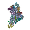
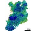
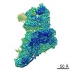
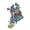
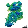
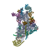
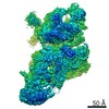
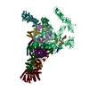
| Title | Structure of the small subunit of the mammalian mitoribosome | |||||||||
 Components Components |
| |||||||||
 Keywords Keywords |  RIBOSOME / TRANSLATION / MITOCHONDRIA / MAMMALIAN 55S MITORIBOSOME / MAMMALIAN 55S MITOCHONDRIAL RIBOSOME / 28S SMALL SUBUNIT / MRNA / TRNA / DECODING CENTER / CRYO-EM / SINGLE PARTICLE ANALYSIS RIBOSOME / TRANSLATION / MITOCHONDRIA / MAMMALIAN 55S MITORIBOSOME / MAMMALIAN 55S MITOCHONDRIAL RIBOSOME / 28S SMALL SUBUNIT / MRNA / TRNA / DECODING CENTER / CRYO-EM / SINGLE PARTICLE ANALYSIS | |||||||||
| Function / homology |  Function and homology information Function and homology informationMitochondrial translation elongation / Mitochondrial translation termination / mitochondrial ribosome assembly / mitochondrial small ribosomal subunit / mitochondrial ribosome / mitochondrial translation / ribosomal small subunit binding / organelle membrane / small ribosomal subunit rRNA binding / regulation of translation ...Mitochondrial translation elongation / Mitochondrial translation termination / mitochondrial ribosome assembly / mitochondrial small ribosomal subunit / mitochondrial ribosome / mitochondrial translation / ribosomal small subunit binding / organelle membrane / small ribosomal subunit rRNA binding / regulation of translation / cell junction / small ribosomal subunit / rRNA binding / ribosome / structural constituent of ribosome / translation / ribonucleoprotein complex / apoptotic process / mitochondrion / RNA binding / nucleoplasm / cytoplasmSimilarity search - Function | |||||||||
| Biological species |  Sus scrofa (pig) Sus scrofa (pig) | |||||||||
| Method | ELECTRON MICROSCOPY / single particle reconstruction / cryo EM / Resolution: 3.6 Å | |||||||||
 Authors Authors | Greber, B.J. / Bieri, P. / Leibundgut, M. / Leitner, A. / Aebersold, R. / Boehringer, D. / Ban, N. | |||||||||
 Citation Citation | Journal: Science / Year: 2015 Title: Ribosome. The complete structure of the 55S mammalian mitochondrial ribosome. Authors: Basil J Greber / Philipp Bieri / Marc Leibundgut / Alexander Leitner / Ruedi Aebersold / Daniel Boehringer / Nenad Ban /  Abstract: Mammalian mitochondrial ribosomes (mitoribosomes) synthesize mitochondrially encoded membrane proteins that are critical for mitochondrial function. Here we present the complete atomic structure of ...Mammalian mitochondrial ribosomes (mitoribosomes) synthesize mitochondrially encoded membrane proteins that are critical for mitochondrial function. Here we present the complete atomic structure of the porcine 55S mitoribosome at 3.8 angstrom resolution by cryo-electron microscopy and chemical cross-linking/mass spectrometry. The structure of the 28S subunit in the complex was resolved at 3.6 angstrom resolution by focused alignment, which allowed building of a detailed atomic structure including all of its 15 mitoribosomal-specific proteins. The structure reveals the intersubunit contacts in the 55S mitoribosome, the molecular architecture of the mitoribosomal messenger RNA (mRNA) binding channel and its interaction with transfer RNAs, and provides insight into the highly specialized mechanism of mRNA recruitment to the 28S subunit. Furthermore, the structure contributes to a mechanistic understanding of aminoglycoside ototoxicity. | |||||||||
| History |
| |||||||||
| Remark 700 | SHEET DETERMINATION METHOD: DSSP THE SHEETS PRESENTED AS "LA" IN EACH CHAIN ON SHEET RECORDS BELOW ... SHEET DETERMINATION METHOD: DSSP THE SHEETS PRESENTED AS "LA" IN EACH CHAIN ON SHEET RECORDS BELOW IS ACTUALLY AN 5-STRANDED BARREL THIS IS REPRESENTED BY A 6-STRANDED SHEET IN WHICH THE FIRST AND LAST STRANDS ARE IDENTICAL. THE SHEETS PRESENTED AS "QA" IN EACH CHAIN ON SHEET RECORDS BELOW IS ACTUALLY AN 5-STRANDED BARREL THIS IS REPRESENTED BY A 6-STRANDED SHEET IN WHICH THE FIRST AND LAST STRANDS ARE IDENTICAL. |
- Structure visualization
| Movie |
Movie viewer |
|---|---|
| Structure viewer | Molecule: MolmilJmol/JSmol |
- Downloads & links
-Download
| PDBx/mmCIF format | 5aj3.cif.gz | 1.7 MB | Display | PDBx/mmCIF format |
|---|---|---|---|---|
| PDB format | pdb5aj3.ent.gz | 1.3 MB | Display | PDB format |
| PDBx/mmJSON format | 5aj3.json.gz | Tree view | PDBx/mmJSON format | |
| Others |  Other downloads Other downloads |
-Validation report
| Arichive directory | https://data.pdbj.org/pub/pdb/validation_reports/aj/5aj3ftp://data.pdbj.org/pub/pdb/validation_reports/aj/5aj3 | HTTPS FTP |
|---|
-Related structure data
| Related structure data | 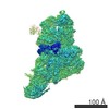 2913MC 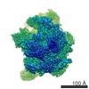 2914C 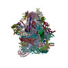 5aj4C M: map data used to model this data C: citing same article ( |
|---|---|
| Similar structure data |


-Links
 PDBj
PDBj


- Assembly
| Deposited unit | 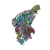
|
|---|---|
| 1 |
|
-Components
-RNA chain , 3 types, 4 molecules AVYX
| #1: RNA chain | Mass: 308989.062 Da / Num. of mol.: 1 / Source method: isolated from a natural source / Source: (natural) Sus scrofa / Organ: LIVER / References: GenBank: 223972359 | ||
|---|---|---|---|
| #18: RNA chain | Mass: 17744.771 Da / Num. of mol.: 2 / Source method: isolated from a natural source Details: MIXTURE OF DIFFERENT MITOCHONDRIAL TRNA SPECIES, BUILT AS POLY-PYRIMIDINE AND POLY-PURINE Source: (natural) Sus scrofa / Organ: LIVER#19: RNA chain | | Messenger RNAMass: 3545.419 Da / Num. of mol.: 1 / Source method: isolated from a natural source Details: MIXTURE OF DIFFERENT MITOCHONDRIAL MRNA SPECIES, BUILT AS POLY-PYRIMIDINE Source: (natural) Sus scrofa / Organ: LIVER |
+MITORIBOSOMAL PROTEIN ... , 31 types, 31 molecules BCEFGIJKLNOPQRTUabcdefghijkmnop
-Protein/peptide , 2 types, 2 molecules sz
| #35: Protein/peptide | Mass: 1379.692 Da / Num. of mol.: 1 / Source method: isolated from a natural source / Source: (natural) Sus scrofa / Organ: LIVER |
|---|---|
| #36: Protein/peptide | Mass: 1464.797 Da / Num. of mol.: 1 / Source method: isolated from a natural source / Source: (natural) Sus scrofa / Organ: LIVER |
-Non-polymers , 4 types, 266 molecules 


| #37: Chemical | ChemComp-MG /  Mass: 24.305 Da / Num. of mol.: 144 / Source method: obtained synthetically / Formula: Mg Mass: 24.305 Da / Num. of mol.: 144 / Source method: obtained synthetically / Formula: Mg#38: Chemical |  Mass: 65.409 Da / Num. of mol.: 3 / Source method: obtained synthetically / Formula: Zn Mass: 65.409 Da / Num. of mol.: 3 / Source method: obtained synthetically / Formula: Zn#39: Chemical | ChemComp-GDP / | Guanosine diphosphate Type: RNA linking / Mass: 443.201 Da / Num. of mol.: 1 / Source method: obtained synthetically / Formula: C10H15N5O11P2 / Comment: GDP, energy-carrying molecule*YM Type: RNA linking / Mass: 443.201 Da / Num. of mol.: 1 / Source method: obtained synthetically / Formula: C10H15N5O11P2 / Comment: GDP, energy-carrying molecule*YM#40: Water | ChemComp-HOH / | WaterMass: 18.015 Da / Num. of mol.: 118 / Source method: isolated from a natural source / Formula: H2O |
|---|
-Details
| Sequence details | CHAIN F GB EW168165.2 CHAIN I GB NP_001231482.1 CHAIN J GB AK233895.1 CHAIN L GB AK394439.1 CHAIN N ...CHAIN F GB EW168165.2 CHAIN I GB NP_001231482.1 CHAIN J GB AK233895.1 CHAIN L GB AK394439.1 CHAIN N GB FD598185.1 CHAIN O GB AK343256.1 CHAIN R GB HX217955.1 CHAIN T UNP I3LNJ0 CHAIN a GB AK348087.1 CHAIN b GB XP_005669015.1 CHAIN c GB AK346624.1 CHAIN e GB XP_003134081.1 CHAIN f GB DN116920.1 CHAIN g GB XP_003361167.1 CHAIN j GB NP_001231761.1 CHAIN n GB AK231191.1 RESIDUES 251-266 OF uS9m (CHAIN I) BUILT AS UNK RESIDUES 309-356 OF mS22 (CHAIN a) BUILT AS UNK PPR FOLD OF mS27 (CHAIN e) BUILT AS UNK RESIDUES 52-69 OF mS29 (CHAIN g) BUILT AS UNK RESIDUES 152-158 OF mS34 (CHAIN j) BUILT AS UNK N-TERMINAL SEQUENCE OF mS38 (CHAIN n) IS MISSING PPR FOLD OF mS39 (CHAIN o) BUILT AS UNK ALL RESIDUES OF CHAINS s,z (UNASSIGNED |
|---|
-Experimental details
-Experiment
| Experiment | Method: ELECTRON MICROSCOPY |
|---|---|
| EM experiment | Aggregation state: PARTICLE / 3D reconstruction method: single particle reconstruction |
- Sample preparation
| Component | Name: SUS SCROFA 55S MITOCHONDRIAL RIBOSOME / Type: RIBOSOME Details: QUANTIFOIL HOLEY CARBON GRIDS WERE COATED WITH A THIN CARBON FILM |
|---|---|
| Buffer solution | Name: 20 MM HEPES-KOH, 50 MM KCL, 40 MM MGCL2, 1 MM DTT / pH: 7.4 / Details: 20 MM HEPES-KOH, 50 MM KCL, 40 MM MGCL2, 1 MM DTT |
| Specimen | Embedding applied: NO / Shadowing applied: NO / Staining applied: NO / Vitrification applied: YES |
| Specimen support | Details: CARBON |
| Vitrification | Instrument: HOMEMADE PLUNGER / Cryogen name: ETHANE-PROPANE / Details: MIXTURE OF LIQUID ETHANE AND PROPANE |
- Electron microscopy imaging
| Experimental equipment |  Model: Titan Krios / Image courtesy: FEI Company |
|---|---|
| Microscopy | Model: FEI TITAN KRIOS / Date: May 30, 2014 Details: IMAGES WERE ACQUIRED IN 2 SESSIONS ON A FEI TITAN KRIOS IN MAY 2014 |
| Electron gun | Electron source: FIELD EMISSION GUN / Accelerating voltage: 300 kV / Illumination mode: FLOOD BEAM |
| Electron lens | Mode: BRIGHT FIELDBright-field microscopy / Nominal magnification: 59000 X / Calibrated magnification: 100000 X / Nominal defocus max: 3000 nm / Nominal defocus min: 800 nm / Cs: 2.7 mm |
| Specimen holder | Temperature: 85 K |
| Image recording | Electron dose: 20 e/Å2 / Film or detector model: FEI FALCON II (4k x 4k) |
| Radiation wavelength | Relative weight: 1 |
- Processing
| EM software |
| ||||||||||||||||||||||||
|---|---|---|---|---|---|---|---|---|---|---|---|---|---|---|---|---|---|---|---|---|---|---|---|---|---|
| CTF correction | Details: PER DETECTOR FRAME | ||||||||||||||||||||||||
| Symmetry | Point symmetry: C1 (asymmetric) | ||||||||||||||||||||||||
| 3D reconstruction | Method: MAXIMUM LIKELIHOOD BASED REFINEMENT IMPLEMENTED IN RELION Resolution: 3.6 Å / Num. of particles: 78783 / Actual pixel size: 1.39 Å Details: FOR VISUALIZATION PURPOSES THE FINAL MAP WAS FILTERED AND AMPLITUDE CORRECTED IN RELION Symmetry type: POINT | ||||||||||||||||||||||||
| Atomic model building | Protocol: OTHER / Space: REAL / Details: REFINEMENT PROTOCOL--HIGH RESOLUTION CRYO-EM | ||||||||||||||||||||||||
| Refinement | Highest resolution: 3.6 Å | ||||||||||||||||||||||||
| Refinement step | Cycle: LAST / Highest resolution: 3.6 Å
|
