 Movie
Movie Controller


+ Open data

- Basic information
| Entry | Database: PDB / ID: 4v8l | ||||||||||||
|---|---|---|---|---|---|---|---|---|---|---|---|---|---|
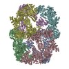
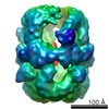
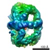
| Title | Cryo-EM Structure of the Mycobacterial Fatty Acid Synthase | ||||||||||||
 Components Components | FATTY ACID SYNTHASE | ||||||||||||
 Keywords Keywords | TRANSFERASE / MYCOLIC ACID BIOSYNTHESIS / MULTIFUNCTIONAL ENZYME / SUBSTRATE CHANNELING | ||||||||||||
| Function / homology |  Function and homology informationfatty acid synthase complex / enoyl-[acyl-carrier-protein] reductase (NADH) activity / fatty acid biosynthetic process / hydrolase activity Function and homology informationfatty acid synthase complex / enoyl-[acyl-carrier-protein] reductase (NADH) activity / fatty acid biosynthetic process / hydrolase activitySimilarity search - Function | ||||||||||||
| Biological species |  MYCOBACTERIUM SMEGMATIS (bacteria) MYCOBACTERIUM SMEGMATIS (bacteria) | ||||||||||||
| Method | ELECTRON MICROSCOPY / single particle reconstruction / cryo EM / Resolution: 7.5 Å | ||||||||||||
 Authors Authors | Boehringer, D. / Ban, N. / Leibundgut, M. | ||||||||||||
 Citation Citation | Journal: J Mol Biol / Year: 2013 Title: 7.5-Å cryo-em structure of the mycobacterial fatty acid synthase. Authors: Daniel Boehringer / Nenad Ban / Marc Leibundgut /  Abstract: The mycobacterial fatty acid synthase (FAS) complex is a giant 2.0-MDa α(6) homohexameric multifunctional enzyme that catalyzes synthesis of fatty acid precursors of mycolic acids, which are major ...The mycobacterial fatty acid synthase (FAS) complex is a giant 2.0-MDa α(6) homohexameric multifunctional enzyme that catalyzes synthesis of fatty acid precursors of mycolic acids, which are major components of the cell wall in Mycobacteria and play an important role in pathogenicity. Here, we present a three-dimensional reconstruction of the Mycobacterium smegmatis FAS complex at 7.5Å, highly homologous to the Mycobacterium tuberculosis multienzyme, by cryo-electron microscopy. Based on the obtained structural data, which allowed us to identify secondary-structure elements, and sequence homology with the fungal FAS, we generated an accurate architectural model of the complex. The FAS system from Mycobacteria resembles a minimized version of the fungal FAS with much larger openings in the reaction chambers. These architectural features of the mycobacterial FAS may be important for the interaction with mycolic acid processing and condensing enzymes that further modify the precursors produced by FAS and for autoactivation of the FAS complex. | ||||||||||||
| History |
| ||||||||||||
| Remark 700 | SHEET DETERMINATION METHOD: DSSP THE SHEETS PRESENTED AS "DD" IN EACH CHAIN ON SHEET RECORDS BELOW ... SHEET DETERMINATION METHOD: DSSP THE SHEETS PRESENTED AS "DD" IN EACH CHAIN ON SHEET RECORDS BELOW IS ACTUALLY AN 8-STRANDED BARREL THIS IS REPRESENTED BY A 9-STRANDED SHEET IN WHICH THE FIRST AND LAST STRANDS ARE IDENTICAL. THE SHEETS PRESENTED AS "ED" IN EACH CHAIN ON SHEET RECORDS BELOW IS ACTUALLY AN 8-STRANDED BARREL THIS IS REPRESENTED BY A 9-STRANDED SHEET IN WHICH THE FIRST AND LAST STRANDS ARE IDENTICAL. THE SHEETS PRESENTED AS "FD" IN EACH CHAIN ON SHEET RECORDS BELOW IS ACTUALLY AN 8-STRANDED BARREL THIS IS REPRESENTED BY A 9-STRANDED SHEET IN WHICH THE FIRST AND LAST STRANDS ARE IDENTICAL. |
- Structure visualization
| Movie |
Movie viewer |
|---|---|
| Structure viewer | Molecule: MolmilJmol/JSmol |
- Downloads & links
-Download
| PDBx/mmCIF format | 4v8l.cif.gz | 2.6 MB | Display | PDBx/mmCIF format |
|---|---|---|---|---|
| PDB format | pdb4v8l.ent.gz | Display | PDB format | |
| PDBx/mmJSON format | 4v8l.json.gz | Tree view | PDBx/mmJSON format | |
| Others |  Other downloads Other downloads |
-Validation report
| Arichive directory | https://data.pdbj.org/pub/pdb/validation_reports/v8/4v8lftp://data.pdbj.org/pub/pdb/validation_reports/v8/4v8l | HTTPS FTP |
|---|
-Related structure data
| Related structure data | 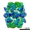 2238MC M: map data used to model this data C: citing same article ( |
|---|---|
| Similar structure data |


-Links
 PDBj
PDBj


- Assembly
| Deposited unit | 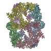
|
|---|---|
| 1 |
|
-Components
| #1: Protein | / TYPE I FATTY ACID SYNTHASE / MYCOBACTERIAL FATTY ACID SYNTHASE I Mass: 325758.250 Da / Num. of mol.: 6 / Source method: isolated from a natural source / Source: (natural) MYCOBACTERIUM SMEGMATIS (bacteria) / Strain: MC2 155 / References: UniProt: A0R1H7#2: Chemical | ChemComp-FMN / Flavin mononucleotide  Mass: 456.344 Da / Num. of mol.: 6 / Source method: obtained synthetically / Formula: C17H21N4O9P Mass: 456.344 Da / Num. of mol.: 6 / Source method: obtained synthetically / Formula: C17H21N4O9P |
|---|
-Experimental details
-Experiment
| Experiment | Method: ELECTRON MICROSCOPY |
|---|---|
| EM experiment | Aggregation state: PARTICLE / 3D reconstruction method: single particle reconstruction |
- Sample preparation
| Component | Name: MYCOBACTERIAL FATTY ACID SYNTHASE, FAS I / Type: COMPLEX |
|---|---|
| Buffer solution | Name: 100MM POTASSIUM PHOSPHATE BUFFER PH 7.2, 165 MM NACL, 2MM EDTA, 2MM DTT pH: 7.2 Details: 100MM POTASSIUM PHOSPHATE BUFFER PH 7.2, 165 MM NACL, 2MM EDTA, 2MM DTT |
| Specimen | Conc.: 1.65 mg/ml / Embedding applied: NO / Shadowing applied: NO / Staining applied: NO / Vitrification applied: YES |
| Specimen support | Details: HOLEY CARBON |
| Vitrification | Instrument: HOMEMADE PLUNGER / Cryogen name: ETHANE / Details: VITRIFIED WITH HOMEMADE PLUNGER IN LIQUID ETHANE |
- Electron microscopy imaging
| Experimental equipment |  Model: Titan Krios / Image courtesy: FEI Company |
|---|---|
| Microscopy | Model: FEI TITAN KRIOS / Date: Oct 19, 2012 Details: DATA WERE COLLECTED USING THE AUTOMATED IMAGE ACQUISITION SOFTWARE FEI EPU. |
| Electron gun | Electron source: FIELD EMISSION GUN / Accelerating voltage: 300 kV / Illumination mode: FLOOD BEAM |
| Electron lens | Mode: BRIGHT FIELDBright-field microscopy / Nominal magnification: 59000 X / Calibrated magnification: 100000 X / Nominal defocus max: 5000 nm / Nominal defocus min: 1500 nm / Cs: 2.7 mm |
| Specimen holder | Temperature: 79 K / Tilt angle max: 0 ° |
| Image recording | Electron dose: 20 e/Å2 / Film or detector model: FEI FALCON I (4k x 4k) |
| Image scans | Num. digital images: 1556 |
| Radiation wavelength | Relative weight: 1 |
- Processing
| EM software |
| ||||||||||||||||
|---|---|---|---|---|---|---|---|---|---|---|---|---|---|---|---|---|---|
| CTF correction | Details: EACH IMAGE | ||||||||||||||||
| Symmetry | Point symmetry: D3 (2x3 fold dihedral) | ||||||||||||||||
| 3D reconstruction | Method: COMMON LINES, PROJECTION MATCHING / Resolution: 7.5 Å / Num. of particles: 106884 / Nominal pixel size: 2.37 Å / Actual pixel size: 1.4 Å Details: SUBMISSION BASED ON EXPERIMENTAL DATA FROM EMDB EMD-2238 (DEPOSITION ID: 11255). Symmetry type: POINT | ||||||||||||||||
| Atomic model building | Protocol: RIGID BODY FIT / Space: REAL / Target criteria: Cross-correlation coefficient Details: METHOD--DOMAINS WERE SEPARATELY FITTED AS RIGID BODIES AND MANUALLY ADJUSTED IN O. THE MODEL WAS MINIMIZED WITH PHENIX.PDBTOOLS. REFINEMENT PROTOCOL--X-RAY | ||||||||||||||||
| Atomic model building | PDB-ID: 2UV8 | ||||||||||||||||
| Refinement | Highest resolution: 7.5 Å | ||||||||||||||||
| Refinement step | Cycle: LAST / Highest resolution: 7.5 Å
|

