 Movie
Movie Controller

[English] 日本語
 Yorodumi
- PDB-4cy4: The Cryo-Electron Microscopy Structure of the CorA channel from M... -
+ Open data

- Basic information
| Entry | Database: PDB / ID: 4cy4 | ||||||
|---|---|---|---|---|---|---|---|
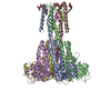
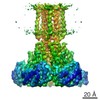
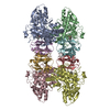
| Title | The Cryo-Electron Microscopy Structure of the CorA channel from Methanocaldococcus jannaschii at 21.6 Angstrom in low magnesium. | ||||||
 Components Components | MAGNESIUM TRANSPORT PROTEIN CORA Magnesium transporter Magnesium transporter | ||||||
 Keywords Keywords | MEMBRANE PROTEIN / MAGNESIUM ION CHANNEL | ||||||
| Function / homology |  Function and homology information Function and homology informationcobalt ion transmembrane transporter activity / magnesium ion transmembrane transporter activity / cobalt ion binding / magnesium ion binding / identical protein binding / plasma membraneSimilarity search - Function | ||||||
| Biological species |   METHANOCALDOCOCCUS JANNASCHII (archaea) METHANOCALDOCOCCUS JANNASCHII (archaea) | ||||||
| Method | ELECTRON MICROSCOPY / single particle reconstruction / cryo EM / Resolution: 21.6 Å | ||||||
| Model type details | CA ATOMS ONLY, CHAIN A, B, C, D, E | ||||||
 Authors Authors | Cleverley, R.M. / Kean, J. / Shintre, C.A. / Baldock, C. / Derrick, J.P. / Ford, R.C. / Prince, S.M. | ||||||
 Citation Citation | Journal: Biochim Biophys Acta / Year: 2015 Title: The Cryo-EM structure of the CorA channel from Methanocaldococcus jannaschii in low magnesium conditions. Authors: Robert M Cleverley / James Kean / Chitra A Shintre / Clair Baldock / Jeremy P Derrick / Robert C Ford / Stephen M Prince /  Abstract: CorA channels are responsible for the uptake of essential magnesium ions by bacteria. X-ray crystal structures have been resolved for two full-length CorA channels, each in a non-conducting state ...CorA channels are responsible for the uptake of essential magnesium ions by bacteria. X-ray crystal structures have been resolved for two full-length CorA channels, each in a non-conducting state with magnesium ions bound to the protein: These structures reveal a homo-pentameric quaternary structure with approximate 5-fold rotational symmetry about a central pore axis. We report the structure of the detergent solubilized Methanocaldococcus jannaschii CorA channel determined by Cryo-Electron Microscopy and Single Particle Averaging, supported by Small Angle X-ray Scattering and X-ray crystallography. This structure also shows a pentameric channel but with a highly asymmetric domain structure. The asymmetry of the domains includes differential separations between the trans-membrane segments, which reflects mechanical coupling of the cytoplasmic domain to the trans-membrane domain. This structure therefore reveals an important aspect of the gating mechanism of CorA channels by providing an indication of how the absence of magnesium ions leads to major structural changes. | ||||||
| History |
|
- Structure visualization
| Movie |
Movie viewer |
|---|---|
| Structure viewer | Molecule: MolmilJmol/JSmol |
- Downloads & links
-Download
| PDBx/mmCIF format | 4cy4.cif.gz | 55 KB | Display | PDBx/mmCIF format |
|---|---|---|---|---|
| PDB format | pdb4cy4.ent.gz | 37.5 KB | Display | PDB format |
| PDBx/mmJSON format | 4cy4.json.gz | Tree view | PDBx/mmJSON format | |
| Others |  Other downloads Other downloads |
-Validation report
| Arichive directory | https://data.pdbj.org/pub/pdb/validation_reports/cy/4cy4ftp://data.pdbj.org/pub/pdb/validation_reports/cy/4cy4 | HTTPS FTP |
|---|
-Related structure data
| Related structure data |  2626MC M: map data used to model this data C: citing same article ( |
|---|---|
| Similar structure data |


-Links
 PDBj
PDBj- Assembly
| Deposited unit | 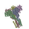
|
|---|---|
| 1 |
|
-Components
| #1: Protein | Magnesium transporter / Coordinate model: Cα atoms only Mass: 37182.586 Da / Num. of mol.: 5 Source method: isolated from a genetically manipulated source Details: RECOMBINANTLY OVER-EXPRESSED PROTEIN, SOLUBILIZED FROM MEMBRANE FRACTION. PURIFIED BY AFFINITY (HIS-TAG), SIZE EXCLUSION CHROMATOGRAPHY. PROTEOLYTIC TAG CLEAVEAGE. Source: (gene. exp.) METHANOCALDOCOCCUS JANNASCHII (archaea)Description: PCR FROM GENOMIC DNA / Plasmid: POPINF / Production host:  ESCHERICHIA COLI (E. coli) / Strain (production host): BL21 / Variant (production host): STAR / References: UniProt: Q58439 ESCHERICHIA COLI (E. coli) / Strain (production host): BL21 / Variant (production host): STAR / References: UniProt: Q58439 |
|---|
-Experimental details
-Experiment
| Experiment | Method: ELECTRON MICROSCOPY |
|---|---|
| EM experiment | Aggregation state: PARTICLE / 3D reconstruction method: single particle reconstruction |
- Sample preparation
| Component | Name: CORA CHANNEL FROM METHANOCALDOCOCCUS JANNASCHII / Type: COMPLEX |
|---|---|
| Buffer solution | Name: 200MM NACL, 20MM TRIS/HCL, 0.04% DODECYLMALTOSIDE ( DDM) pH: 8 Details: 200MM NACL, 20MM TRIS/HCL, 0.04% DODECYLMALTOSIDE ( DDM) |
| Specimen | Conc.: 1 mg/ml / Embedding applied: NO / Shadowing applied: NO / Staining applied: NO / Vitrification applied: YES |
| Specimen support | Details: HOLEY CARBON |
| Vitrification | Instrument: FEI VITROBOT MARK I / Cryogen name: ETHANE / Details: LIQUID ETHANE |
- Electron microscopy imaging
| Experimental equipment |  Model: Tecnai F20 / Image courtesy: FEI Company |
|---|---|
| Microscopy | Model: FEI TECNAI F20 / Date: Jun 5, 2009 / Details: LOW DOSE MODE |
| Electron gun | Electron source: FIELD EMISSION GUN / Accelerating voltage: 200 kV / Illumination mode: FLOOD BEAM |
| Electron lens | Mode: BRIGHT FIELDBright-field microscopy / Nominal defocus max: 4800 nm / Nominal defocus min: 3300 nm |
| Specimen holder | Temperature: 100 K |
| Image recording | Film or detector model: GATAN ULTRASCAN 4000 (4k x 4k) |
| Image scans | Num. digital images: 28 |
- Processing
| EM software |
| ||||||||||||
|---|---|---|---|---|---|---|---|---|---|---|---|---|---|
| CTF correction | Details: WITH REFERENCE TO X-RAY SCATTERING CURVE | ||||||||||||
| Symmetry | Point symmetry: C1 (asymmetric) | ||||||||||||
| 3D reconstruction | Method: 3D RECONSTRUCTION FROM RANDOMLY ORIENTED PARTICLE IMAGES Resolution: 21.6 Å / Num. of particles: 44064 / Nominal pixel size: 3.5 Å / Actual pixel size: 3.5 Å Details: THE HIGH-RES MJCORA PDB 4EV6 STRUCTURE WAS DOCKED AS AN UNMODIFIED RIGID BODY. THERE ARE DIFFERENCES IN THE CRYO- EM AND CRYSTAL STRUCTURE DUE TO DIFFERENCES IN SOLUTION CONDITIONS ...Details: THE HIGH-RES MJCORA PDB 4EV6 STRUCTURE WAS DOCKED AS AN UNMODIFIED RIGID BODY. THERE ARE DIFFERENCES IN THE CRYO- EM AND CRYSTAL STRUCTURE DUE TO DIFFERENCES IN SOLUTION CONDITIONS SPECIFICALLY MG ION CONCENTRATION. SUBMISSION BASED ON EXPERIMENTAL DATA FROM EMDB EMD-2626. (DEPOSITION ID: 12440). Symmetry type: POINT | ||||||||||||
| Atomic model building | Protocol: RIGID BODY FIT / Space: REAL / Target criteria: Cross-correlation coefficient Details: METHOD--RIGID BODY REFINEMENT PROTOCOL--DOCKING OF X-RAY CRYSTAL STRUCTURE | ||||||||||||
| Atomic model building | PDB-ID: 4EV6 | ||||||||||||
| Refinement | Highest resolution: 21.6 Å | ||||||||||||
| Refinement step | Cycle: LAST / Highest resolution: 21.6 Å
|

