Movie
Movie Controller

[English] 日本語
 Yorodumi
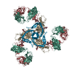
- PDB-4cc8: Pre-fusion structure of trimeric HIV-1 envelope glycoprotein dete... -
+ Open data

- Basic information
| Entry | Database: PDB / ID: 4cc8 | |||||||||
|---|---|---|---|---|---|---|---|---|---|---|
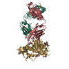
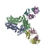
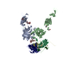
| Title | Pre-fusion structure of trimeric HIV-1 envelope glycoprotein determined by cryo-electron microscopy | |||||||||
 Components Components |
| |||||||||
 Keywords Keywords |  VIRAL PROTEIN/IMMUNE SYSTEM / VIRAL PROTEIN-IMMUNE SYSTEM COMPLEX / PRE-FUSION STATE VIRAL PROTEIN/IMMUNE SYSTEM / VIRAL PROTEIN-IMMUNE SYSTEM COMPLEX / PRE-FUSION STATE | |||||||||
| Biological species |   HUMAN IMMUNODEFICIENCY VIRUS 1 HUMAN IMMUNODEFICIENCY VIRUS 1 HOMO SAPIENS (human) HOMO SAPIENS (human) | |||||||||
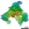
| Method | ELECTRON MICROSCOPY / single particle reconstruction / cryo EM / Resolution: 6 Å | |||||||||
 Authors Authors | Bartesaghi, A. / Merk, A. / Borgnia, M.J. / Milne, J.L.S. / Subramaniam, S. | |||||||||
 Citation Citation | Journal: Nat Struct Mol Biol / Year: 2013 Title: Prefusion structure of trimeric HIV-1 envelope glycoprotein determined by cryo-electron microscopy. Authors: Alberto Bartesaghi / Alan Merk / Mario J Borgnia / Jacqueline L S Milne / Sriram Subramaniam /  Abstract: The activation of trimeric HIV-1 envelope glycoprotein (Env) by its binding to the cell-surface receptor CD4 and co-receptors (CCR5 or CXCR4) represents the first of a series of events that lead to ...The activation of trimeric HIV-1 envelope glycoprotein (Env) by its binding to the cell-surface receptor CD4 and co-receptors (CCR5 or CXCR4) represents the first of a series of events that lead to fusion between viral and target-cell membranes. Here, we present the cryo-EM structure, at subnanometer resolution (~6 Å at 0.143 FSC), of the 'closed', prefusion state of trimeric HIV-1 Env complexed to the broadly neutralizing antibody VRC03. We show that three gp41 helices at the core of the trimer serve as an anchor around which the rest of Env is reorganized upon activation to the 'open' quaternary conformation. The architecture of trimeric HIV-1 Env in the prefusion state and in the activated intermediate state resembles the corresponding states of influenza hemagglutinin trimers, thus providing direct evidence for the similarity in entry mechanisms used by HIV-1, influenza and related enveloped viruses. | |||||||||
| History |
|
- Structure visualization
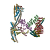
| Movie |
 Movie viewer Movie viewer |
|---|---|
| Structure viewer | Molecule: MolmilJmol/JSmol |
- Downloads & links
-Download
| PDBx/mmCIF format | 4cc8.cif.gz | 1.2 MB | Display | PDBx/mmCIF format |
|---|---|---|---|---|
| PDB format | pdb4cc8.ent.gz | 1.1 MB | Display | PDB format |
| PDBx/mmJSON format | 4cc8.json.gz | Tree view | PDBx/mmJSON format | |
| Others |  Other downloads Other downloads |
-Validation report
| Arichive directory | https://data.pdbj.org/pub/pdb/validation_reports/cc/4cc8ftp://data.pdbj.org/pub/pdb/validation_reports/cc/4cc8 | HTTPS FTP |
|---|
-Related structure data
| Related structure data |  2484MC M: map data used to model this data C: citing same article ( |
|---|---|
| Similar structure data | |
| EM raw data | EMPIAR-10004 (Title: Pre-fusion structure of trimeric HIV-1 envelope glycoprotein determined by cryo-electron microscopy Data size: 117.8 Data #1: HIV-1 envelope glycoprotein micrographs [micrographs - single frame]) |
-Links
 PDBj
PDBj


- Assembly
| Deposited unit | 
|
|---|---|
| 1 | 
|
-Components
| #1: Antibody | Mass: 2656.265 Da / Num. of mol.: 3 Source method: isolated from a genetically manipulated source Details: COMPLETE ECTODOMAIN OF HIV-1 ENV FROM THE CLADE A STRAIN KNH1144 INLCUDING RESIDUES IN THE MEMBRANE PROXIMAL EXTERNAL REGION WITH THE FOLLOWING RESIDUE SUBSTITUTIONS A662E, S668N, AND S676T Source: (gene. exp.) HUMAN IMMUNODEFICIENCY VIRUS 1 / Variant: HIV-1 ISOLATE 00KE_KNH1144 / Plasmid: SOSIP-PPI4, FURIN-PCDNA3.1 / Cell line (production host): HEK293 / Production host: HOMO SAPIENS (human)#2: Antibody | Envelope glycoprotein GP120Mass: 29293.975 Da / Num. of mol.: 3 Source method: isolated from a genetically manipulated source Source: (gene. exp.) HUMAN IMMUNODEFICIENCY VIRUS 1 / Variant: HIV-1 ISOLATE 00KE_KNH1144 / Plasmid: SOSIP-PPI4, FURIN-PCDNA3.1 / Cell line (production host): HEK293 / Production host: HOMO SAPIENS (human)#3: Antibody | Mass: 25294.525 Da / Num. of mol.: 3 Source method: isolated from a genetically manipulated source Source: (gene. exp.) HOMO SAPIENS (human) / Cell line (production host): HEK293 / Production host: HOMO SAPIENS (human)#4: Antibody | Mass: 23146.795 Da / Num. of mol.: 3 Source method: isolated from a genetically manipulated source Source: (gene. exp.) HOMO SAPIENS (human) / Cell line (production host): HEK293 / Production host: HOMO SAPIENS (human)Sequence details | THE GP41 COMPONENT (CHAINS A, B, C) INCLUDES UNIPROT Q3ZLH6 RESIDUES 512-681 WITH THE FOLLOWING ...THE GP41 COMPONENT (CHAINS A, B, C) INCLUDES UNIPROT Q3ZLH6 RESIDUES 512-681 WITH THE FOLLOWING SUBSTITUTI | |
|---|
-Experimental details
-Experiment
| Experiment | Method: ELECTRON MICROSCOPY |
|---|---|
| EM experiment | Aggregation state: PARTICLE / 3D reconstruction method: single particle reconstruction |
- Sample preparation
| Component | Name: COMPLEX OF KNH1144 SOSIP GP140 TRIMER WITH VRC03 FAB. / Type: COMPLEX |
|---|---|
| Buffer solution | Name: TNE BUFFER (10 MM TRIS, 150 MM NACL, 1 MM EDTA) / pH: 7.5 / Details: TNE BUFFER (10 MM TRIS, 150 MM NACL, 1 MM EDTA) |
| Specimen | Conc.: 0.65 mg/ml / Embedding applied: NO / Shadowing applied: NO / Staining applied: NO / Vitrification applied: YES |
| Specimen support | Details: HOLEY CARBON |
| Vitrification | Instrument: FEI VITROBOT MARK IV / Cryogen name: ETHANE Details: BLOT FOR 6 SECONDS, BLOT OFFSET OF -2, PLUNGE INTO AN ETHANE SLURRY COOLED BY LIQUID NITROGEN |
- Electron microscopy imaging
| Experimental equipment |  Model: Titan Krios / Image courtesy: FEI Company |
|---|---|
| Microscopy | Model: FEI TITAN KRIOS / Date: Aug 9, 2011 |
| Electron gun | Electron source: FIELD EMISSION GUN / Accelerating voltage: 80 kV / Illumination mode: FLOOD BEAM |
| Electron lens | Mode: BRIGHT FIELDBright-field microscopy / Nominal magnification: 75000 X / Nominal defocus max: 2500 nm / Nominal defocus min: 1500 nm / Cs: 2.7 mm |
| Specimen holder | Temperature: 80 K |
| Image recording | Electron dose: 10 e/Å2 / Film or detector model: GATAN ULTRASCAN 4000 (4k x 4k) |
| Image scans | Num. digital images: 2000 |
| Radiation wavelength | Relative weight: 1 |
- Processing
| EM software |
| |||||||||||||||
|---|---|---|---|---|---|---|---|---|---|---|---|---|---|---|---|---|
| Symmetry | Point symmetry: C3 (3 fold cyclic) | |||||||||||||||
| 3D reconstruction | Resolution: 6 Å / Num. of particles: 88125 / Nominal pixel size: 1.08 Å Details: 3SE8, CHAIN G AND CHAINS H, L FITTED AS TWO SEPARATE RIGID BODIES INTO MAP. ONLY HELIX RESIDUES 92-122 OF CHAIN B FITTED BY HAND INTO MAP. SUBMISSION BASED ON EXPERIMENTAL DATA FROM EMDB EMD- ...Details: 3SE8, CHAIN G AND CHAINS H, L FITTED AS TWO SEPARATE RIGID BODIES INTO MAP. ONLY HELIX RESIDUES 92-122 OF CHAIN B FITTED BY HAND INTO MAP. SUBMISSION BASED ON EXPERIMENTAL DATA FROM EMDB EMD-2484. (DEPOSITION ID: 12023). Symmetry type: POINT | |||||||||||||||
| Atomic model building | Protocol: RIGID BODY FIT / Space: RECIPROCAL / Details: REFINEMENT PROTOCOL--RIGID BODY | |||||||||||||||
| Atomic model building |
| |||||||||||||||
| Refinement | Highest resolution: 6 Å | |||||||||||||||
| Refinement step | Cycle: LAST / Highest resolution: 6 Å
|