 Movie
Movie Controller

[English] 日本語
 Yorodumi
- PDB-4bx4: Fitting of the bacteriophage Phi8 P1 capsid protein into cryo-EM ... -
+ Open data

- Basic information
| Entry | Database: PDB / ID: 4bx4 | ||||||
|---|---|---|---|---|---|---|---|
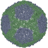
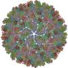
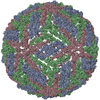
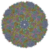
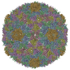
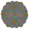
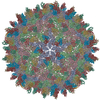
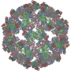
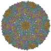
| Title | Fitting of the bacteriophage Phi8 P1 capsid protein into cryo-EM density | ||||||
 Components Components | P1 | ||||||
 Keywords Keywords |  VIRUS / BACTERIOPHAGE VIRUS / BACTERIOPHAGE | ||||||
| Function / homology | : / Major inner capsid protein P1 / identical protein binding / p1 Function and homology information Function and homology information | ||||||
| Biological species |  PSEUDOMONAS PHAGE PHI8 (bacteriophage) PSEUDOMONAS PHAGE PHI8 (bacteriophage) | ||||||
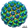
| Method | ELECTRON MICROSCOPY / single particle reconstruction / cryo EM / Resolution: 8.7 Å | ||||||
 Authors Authors | El Omari, K. / Sutton, G. / Ravantti, J.J. / Zhang, H. / Walter, T.S. / Grimes, J.M. / Bamford, D.H. / Stuart, D.I. / Mancini, E.J. | ||||||
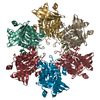
 Citation Citation | Journal: Structure / Year: 2013 Title: Plate tectonics of virus shell assembly and reorganization in phage φ8, a distant relative of mammalian reoviruses. Authors: Kamel El Omari / Geoff Sutton / Janne J Ravantti / Hanwen Zhang / Thomas S Walter / Jonathan M Grimes / Dennis H Bamford / David I Stuart / Erika J Mancini /  Abstract: The hallmark of a virus is its capsid, which harbors the viral genome and is formed from protein subunits, which assemble following precise geometric rules. dsRNA viruses use an unusual protein ...The hallmark of a virus is its capsid, which harbors the viral genome and is formed from protein subunits, which assemble following precise geometric rules. dsRNA viruses use an unusual protein multiplicity (120 copies) to form their closed capsids. We have determined the atomic structure of the capsid protein (P1) from the dsRNA cystovirus Φ8. In the crystal P1 forms pentamers, very similar in shape to facets of empty procapsids, suggesting an unexpected assembly pathway that proceeds via a pentameric intermediate. Unlike the elongated proteins used by dsRNA mammalian reoviruses, P1 has a compact trapezoid-like shape and a distinct arrangement in the shell, with two near-identical conformers in nonequivalent structural environments. Nevertheless, structural similarity with the analogous protein from the mammalian viruses suggests a common ancestor. The unusual shape of the molecule may facilitate dramatic capsid expansion during phage maturation, allowing P1 to switch interaction interfaces to provide capsid plasticity. | ||||||
| History |
|
- Structure visualization
| Movie |
Movie viewer |
|---|---|
| Structure viewer | Molecule: MolmilJmol/JSmol |
- Downloads & links
-Download
| PDBx/mmCIF format | 4bx4.cif.gz | 253.9 KB | Display | PDBx/mmCIF format |
|---|---|---|---|---|
| PDB format | pdb4bx4.ent.gz | 205.2 KB | Display | PDB format |
| PDBx/mmJSON format | 4bx4.json.gz | Tree view | PDBx/mmJSON format | |
| Others |  Other downloads Other downloads |
-Validation report
| Arichive directory | https://data.pdbj.org/pub/pdb/validation_reports/bx/4bx4ftp://data.pdbj.org/pub/pdb/validation_reports/bx/4bx4 | HTTPS FTP |
|---|
-Related structure data
| Related structure data | 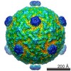 1300M  4btpC M: map data used to model this data C: citing same article ( |
|---|---|
| Similar structure data |
-Links
 PDBj
PDBj- Assembly
| Deposited unit | 
|
|---|---|
| 1 | x 60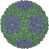
|
| 2 |
|
| 3 | x 5
|
| 4 | x 6
|
| 5 | 
|
| Symmetry | Point symmetry: (Schoenflies symbol: I (icosahedral)) |
| Noncrystallographic symmetry (NCS) | NCS oper: (Code: given / Matrix: (1), |
-Components
| #1: Protein | Mass: 86971.078 Da / Num. of mol.: 2 Source method: isolated from a genetically manipulated source Source: (gene. exp.) PSEUDOMONAS PHAGE PHI8 (bacteriophage) / Plasmid: PDK5 / Production host:  ESCHERICHIA COLI (E. coli) / Strain (production host): BL21(DE3) / References: UniProt: Q9MC13 ESCHERICHIA COLI (E. coli) / Strain (production host): BL21(DE3) / References: UniProt: Q9MC13 |
|---|
-Experimental details
-Experiment
| Experiment | Method: ELECTRON MICROSCOPY |
|---|---|
| EM experiment | Aggregation state: PARTICLE / 3D reconstruction method: single particle reconstruction |
- Sample preparation
| Component | Name: PSEUDOMONAS PHAGE PHI8 / Type: VIRUS |
|---|---|
| Buffer solution | Name: 10 MM POTASSIUM PHOSPHATE PH7.5, 1 MM MGCL2, 50 MM NACL pH: 7.5 Details: 10 MM POTASSIUM PHOSPHATE PH7.5, 1 MM MGCL2, 50 MM NACL |
| Specimen | Embedding applied: NO / Shadowing applied: NO / Staining applied: NO / Vitrification applied: YES |
| Specimen support | Details: OTHER |
| Vitrification | Instrument: HOMEMADE PLUNGER / Cryogen name: ETHANE |
- Electron microscopy imaging
| Experimental equipment |  Model: Tecnai F20 / Image courtesy: FEI Company |
|---|---|
| Microscopy | Model: FEI TECNAI F20 |
| Electron gun | Electron source: FIELD EMISSION GUN / Accelerating voltage: 200 kV / Illumination mode: FLOOD BEAM |
| Electron lens | Mode: BRIGHT FIELDBright-field microscopy / Nominal magnification: 50000 X / Calibrated magnification: 49300 X / Nominal defocus max: 3300 nm / Nominal defocus min: 700 nm / Cs: 2 mm |
| Image recording | Film or detector model: KODAK SO-163 FILM |
| Image scans | Num. digital images: 66 |
- Processing
| EM software |
| ||||||||||||||||||||||||
|---|---|---|---|---|---|---|---|---|---|---|---|---|---|---|---|---|---|---|---|---|---|---|---|---|---|
| Symmetry | Point symmetry: I (icosahedral) | ||||||||||||||||||||||||
| 3D reconstruction | Resolution: 8.7 Å / Num. of particles: 12867 / Details: COORDINATES FITTED TO CRYO-EM MAP EMD-1300 / Symmetry type: POINT | ||||||||||||||||||||||||
| Atomic model building | Protocol: OTHER / Space: REAL / Details: REFINEMENT PROTOCOL--X-RAY | ||||||||||||||||||||||||
| Atomic model building | PDB-ID: 4BTP | ||||||||||||||||||||||||
| Refinement | Highest resolution: 8.7 Å | ||||||||||||||||||||||||
| Refinement step | Cycle: LAST / Highest resolution: 8.7 Å
|
