 Movie
Movie Controller

[English] 日本語
 Yorodumi
- PDB-4au6: Location of the dsRNA-dependent polymerase, VP1, in rotavirus par... -
+ Open data

- Basic information
| Entry | Database: PDB / ID: 4au6 | ||||||
|---|---|---|---|---|---|---|---|
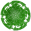
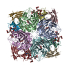
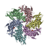
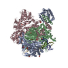
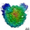
| Title | Location of the dsRNA-dependent polymerase, VP1, in rotavirus particles | ||||||
 Components Components | RNA-DEPENDENT RNA POLYMERASE | ||||||
 Keywords Keywords | VIRAL PROTEIN / DSRNA-DEPENDENT | ||||||
| Function / homology |  Function and homology informationvirion component => GO:0044423 / viral genome replication / RNA-directed RNA polymerase / RNA-dependent RNA polymerase activity / nucleotide binding / DNA-templated transcription / RNA binding Function and homology informationvirion component => GO:0044423 / viral genome replication / RNA-directed RNA polymerase / RNA-dependent RNA polymerase activity / nucleotide binding / DNA-templated transcription / RNA bindingSimilarity search - Function | ||||||
| Biological species |  BOVINE ROTAVIRUS BOVINE ROTAVIRUS | ||||||
| Method | ELECTRON MICROSCOPY / single particle reconstruction / cryo EM / Resolution: 6 Å | ||||||
 Authors Authors | Estrozi, L.F. / Settembre, E.C. / Goret, G. / McClain, B. / Zhang, X. / Chen, J.Z. / Grigorieff, N. / Harrison, S.C. | ||||||
 Citation Citation | Journal: J Mol Biol / Year: 2013 Title: Location of the dsRNA-dependent polymerase, VP1, in rotavirus particles. Authors: Leandro F Estrozi / Ethan C Settembre / Gaël Goret / Brian McClain / Xing Zhang / James Z Chen / Nikolaus Grigorieff / Stephen C Harrison /  Abstract: Double-stranded RNA (dsRNA) viruses transcribe and replicate RNA within an assembled, inner capsid particle; only plus-sense mRNA emerges into the intracellular milieu. During infectious entry of a ...Double-stranded RNA (dsRNA) viruses transcribe and replicate RNA within an assembled, inner capsid particle; only plus-sense mRNA emerges into the intracellular milieu. During infectious entry of a rotavirus particle, the outer layer of its three-layer structure dissociates, delivering the inner double-layered particle (DLP) into the cytosol. DLP structures determined by X-ray crystallography and electron cryomicroscopy (cryoEM) show that the RNA coils uniformly into the particle interior, avoiding a "fivefold hub" of more structured density projecting inward from the VP2 shell of the DLP along each of the twelve 5-fold axes. Analysis of the X-ray crystallographic electron density map suggested that principal contributors to the hub are the N-terminal arms of VP2, but reexamination of the cryoEM map has shown that many features come from a molecule of VP1, randomly occupying five equivalent and partly overlapping positions. We confirm here that the electron density in the X-ray map leads to the same conclusion, and we describe the functional implications of the orientation and position of the polymerase. The exit channel for the nascent transcript directs the nascent transcript toward an opening along the 5-fold axis. The template strand enters from within the particle, and the dsRNA product of the initial replication step exits in a direction tangential to the inner surface of the VP2 shell, allowing it to coil optimally within the DLP. The polymerases of reoviruses appear to have similar positions and functional orientations. | ||||||
| History |
|
- Structure visualization
| Movie |
Movie viewer |
|---|---|
| Structure viewer | Molecule: MolmilJmol/JSmol |
- Downloads & links
-Download
| PDBx/mmCIF format | 4au6.cif.gz | 1.4 MB | Display | PDBx/mmCIF format |
|---|---|---|---|---|
| PDB format | pdb4au6.ent.gz | 889.8 KB | Display | PDB format |
| PDBx/mmJSON format | 4au6.json.gz | Tree view | PDBx/mmJSON format | |
| Others |  Other downloads Other downloads |
-Validation report
| Arichive directory | https://data.pdbj.org/pub/pdb/validation_reports/au/4au6ftp://data.pdbj.org/pub/pdb/validation_reports/au/4au6 | HTTPS FTP |
|---|
-Related structure data
| Related structure data |  2100MC  4f5xC M: map data used to model this data C: citing same article ( |
|---|---|
| Similar structure data |

-Links
 PDBj
PDBj- Assembly
| Deposited unit | 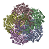
|
|---|---|
| 1 |
|
-Components
| #1: Protein | Mass: 126324.484 Da / Num. of mol.: 5 / Source method: obtained synthetically / Source: (synth.) BOVINE ROTAVIRUSReferences: UniProt: B3IWN8*PLUS, RNA-directed RNA polymeraseSequence details | THIS SEQUENCE IS FITTED FROM SIMIAN ROTAVIRUS, UNIPROT ID 10922 | |
|---|
-Experimental details
-Experiment
| Experiment | Method: ELECTRON MICROSCOPY |
|---|---|
| EM experiment | Aggregation state: PARTICLE / 3D reconstruction method: single particle reconstruction |
- Sample preparation
| Component | Name: ROTAVIRUS DLP / Type: VIRUS |
|---|---|
| Buffer solution | pH: 7.4 |
| Specimen | Conc.: 5 mg/ml / Embedding applied: NO / Shadowing applied: NO / Staining applied: NO / Vitrification applied: YES |
| Specimen support | Details: HOLEY CARBON |
| Vitrification | Instrument: HOMEMADE PLUNGER / Cryogen name: ETHANE / Details: LIQUID ETHANE |
- Electron microscopy imaging
| Experimental equipment |  Model: Tecnai F30 / Image courtesy: FEI Company |
|---|---|
| Microscopy | Model: FEI TECNAI F30 / Date: Jun 1, 2007 |
| Electron gun | Electron source: FIELD EMISSION GUN / Accelerating voltage: 300 kV / Illumination mode: FLOOD BEAM |
| Electron lens | Mode: BRIGHT FIELDBright-field microscopy / Nominal magnification: 59000 X / Calibrated magnification: 56540 X / Nominal defocus max: 3500 nm / Nominal defocus min: 1100 nm / Cs: 2 mm |
| Specimen holder | Temperature: 90 K / Tilt angle max: 0.001 ° / Tilt angle min: 0 ° |
| Image recording | Electron dose: 15 e/Å2 / Film or detector model: KODAK SO-163 FILM |
| Image scans | Num. digital images: 386 |
| Radiation wavelength | Relative weight: 1 |
- Processing
| EM software |
| |||||||||||||||
|---|---|---|---|---|---|---|---|---|---|---|---|---|---|---|---|---|
| CTF correction | Details: INDIVIDUAL PARTICLE PHASE FLIPPING | |||||||||||||||
| Symmetry | Point symmetry: I (icosahedral) | |||||||||||||||
| 3D reconstruction | Method: SYMMETRY-ADAPTED FUNCTIONS / Resolution: 6 Å / Num. of particles: 7000 / Nominal pixel size: 1.18 Å / Actual pixel size: 1.23 Å Details: SUBMISSION BASED ON EXPERIMENTAL DATA FROM EMDB EMD-2100.(DEPOSITION ID: 10810). Symmetry type: POINT | |||||||||||||||
| Atomic model building | Protocol: OTHER / Space: RECIPROCAL / Target criteria: Cross-correlation coefficient / Details: METHOD--URO REFINEMENT PROTOCOL--X-RAY | |||||||||||||||
| Atomic model building | PDB-ID: 2R7O | |||||||||||||||
| Refinement | Highest resolution: 6 Å | |||||||||||||||
| Refinement step | Cycle: LAST / Highest resolution: 6 Å
|

