 Movie
Movie Controller

[English] 日本語
 Yorodumi
- PDB-4adx: The Cryo-EM Structure of the Archaeal 50S Ribosomal Subunit in Co... -
+ Open data

- Basic information
| Entry | Database: PDB / ID: 4adx | ||||||
|---|---|---|---|---|---|---|---|
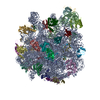
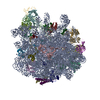
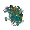
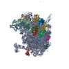
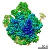
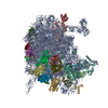
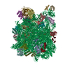
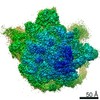
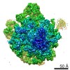
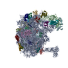
| Title | The Cryo-EM Structure of the Archaeal 50S Ribosomal Subunit in Complex with Initiation Factor 6 | ||||||
 Components Components |
| ||||||
 Keywords Keywords |  RIBOSOME / PROTEIN SYNTHESIS RIBOSOME / PROTEIN SYNTHESIS | ||||||
| Function / homology |  Function and homology informationribonuclease P activity / tRNA 5'-leader removal / translation initiation factor activity / cytosolic ribosome / cytosolic ribosome assembly / large ribosomal subunit rRNA binding / ribosome binding / large ribosomal subunit / ribosome biogenesis / 5S rRNA binding ...ribonuclease P activity / tRNA 5'-leader removal / translation initiation factor activity / cytosolic ribosome / cytosolic ribosome assembly / large ribosomal subunit rRNA binding / ribosome binding / large ribosomal subunit / ribosome biogenesis / 5S rRNA binding / cytosolic large ribosomal subunit / tRNA binding / rRNA binding / ribosome / structural constituent of ribosome / translation / ribonucleoprotein complex / nucleotide binding / DNA repair / DNA binding / RNA binding / zinc ion binding / nucleus / cytoplasm Function and homology informationribonuclease P activity / tRNA 5'-leader removal / translation initiation factor activity / cytosolic ribosome / cytosolic ribosome assembly / large ribosomal subunit rRNA binding / ribosome binding / large ribosomal subunit / ribosome biogenesis / 5S rRNA binding ...ribonuclease P activity / tRNA 5'-leader removal / translation initiation factor activity / cytosolic ribosome / cytosolic ribosome assembly / large ribosomal subunit rRNA binding / ribosome binding / large ribosomal subunit / ribosome biogenesis / 5S rRNA binding / cytosolic large ribosomal subunit / tRNA binding / rRNA binding / ribosome / structural constituent of ribosome / translation / ribonucleoprotein complex / nucleotide binding / DNA repair / DNA binding / RNA binding / zinc ion binding / nucleus / cytoplasmSimilarity search - Function | ||||||
| Biological species |   METHANOTHERMOBACTER THERMAUTOTROPHICUS (archaea) METHANOTHERMOBACTER THERMAUTOTROPHICUS (archaea) | ||||||
| Method | ELECTRON MICROSCOPY / single particle reconstruction / cryo EM / Resolution: 6.6 Å | ||||||
| Model type details | CA ATOMS ONLY, CHAIN Z, H, J, K, L, M, N, O, P, S, V, T, R, X, E, B, C, A, D, F, 3, Q, U, I, W, Y, ...CA ATOMS ONLY, CHAIN Z, H, J, K, L, M, N, O, P, S, V, T, R, X, E, B, C, A, D, F, 3, Q, U, I, W, Y, 1, 2, 4, 5, 6, 7, G ; P ATOMS ONLY, CHAIN 9, 0, 8 | ||||||
 Authors Authors | Greber, B.J. / Boehringer, D. / Godinic-Mikulcic, V. / Crnkovic, A. / Ibba, M. / Weygand-Durasevic, I. / Ban, N. | ||||||
 Citation Citation | Journal: J Mol Biol / Year: 2012 Title: Cryo-EM structure of the archaeal 50S ribosomal subunit in complex with initiation factor 6 and implications for ribosome evolution. Authors: Basil J Greber / Daniel Boehringer / Vlatka Godinic-Mikulcic / Ana Crnkovic / Michael Ibba / Ivana Weygand-Durasevic / Nenad Ban /  Abstract: Translation of mRNA into proteins by the ribosome is universally conserved in all cellular life. The composition and complexity of the translation machinery differ markedly between the three domains ...Translation of mRNA into proteins by the ribosome is universally conserved in all cellular life. The composition and complexity of the translation machinery differ markedly between the three domains of life. Organisms from the domain Archaea show an intermediate level of complexity, sharing several additional components of the translation machinery with eukaryotes that are absent in bacteria. One of these translation factors is initiation factor 6 (IF6), which associates with the large ribosomal subunit. We have reconstructed the 50S ribosomal subunit from the archaeon Methanothermobacter thermautotrophicus in complex with archaeal IF6 at 6.6 Å resolution using cryo-electron microscopy (EM). The structure provides detailed architectural insights into the 50S ribosomal subunit from a methanogenic archaeon through identification of the rRNA expansion segments and ribosomal proteins that are shared between this archaeal ribosome and eukaryotic ribosomes but are mostly absent in bacteria and in some archaeal lineages. Furthermore, the structure reveals that, in spite of highly divergent evolutionary trajectories of the ribosomal particle and the acquisition of novel functions of IF6 in eukaryotes, the molecular binding of IF6 on the ribosome is conserved between eukaryotes and archaea. The structure also provides a snapshot of the reductive evolution of the archaeal ribosome and offers new insights into the evolution of the translation system in archaea. | ||||||
| History |
|
- Structure visualization
| Movie |
Movie viewer |
|---|---|
| Structure viewer | Molecule: MolmilJmol/JSmol |
- Downloads & links
-Download
| PDBx/mmCIF format | 4adx.cif.gz | 274.6 KB | Display | PDBx/mmCIF format |
|---|---|---|---|---|
| PDB format | pdb4adx.ent.gz | 171.2 KB | Display | PDB format |
| PDBx/mmJSON format | 4adx.json.gz | Tree view | PDBx/mmJSON format | |
| Others |  Other downloads Other downloads |
-Validation report
| Arichive directory | https://data.pdbj.org/pub/pdb/validation_reports/ad/4adxftp://data.pdbj.org/pub/pdb/validation_reports/ad/4adx | HTTPS FTP |
|---|
-Related structure data
| Related structure data |  2012MC M: map data used to model this data C: citing same article ( |
|---|---|
| Similar structure data |
-Links
 PDBj
PDBj


- Assembly
| Deposited unit | 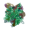
|
|---|---|
| 1 |
|
-Components
-RNA chain , 3 types, 3 molecules 089
| #1: RNA chain | / Coordinate model: P atoms only Mass: 946407.500 Da / Num. of mol.: 1 / Source method: isolated from a natural source Source: (natural) METHANOTHERMOBACTER THERMAUTOTROPHICUS (archaea)Cellular location: CYTOPLASM / Strain: DELTA H |
|---|---|
| #9: RNA chain | Mass: 43052.668 Da / Num. of mol.: 1 / Source method: isolated from a natural source Source: (natural) METHANOTHERMOBACTER THERMAUTOTROPHICUS (archaea)Cellular location: CYTOPLASM / Strain: DELTA H |
| #10: RNA chain | / Coordinate model: P atoms only Mass: 39303.402 Da / Num. of mol.: 1 / Source method: isolated from a natural source Source: (natural) METHANOTHERMOBACTER THERMAUTOTROPHICUS (archaea)Cellular location: CYTOPLASM / Strain: DELTA H |
+Protein , 32 types, 32 molecules 134567ABCDEFGHIJKLMNOPQRSTUVWXYZ
-Protein/peptide , 1 types, 1 molecules 2
| #3: Protein/peptide | Mass: 6133.090 Da / Num. of mol.: 1 / Source method: isolated from a natural source Source: (natural) METHANOTHERMOBACTER THERMAUTOTROPHICUS (archaea)Cellular location: CYTOPLASM / Strain: DELTA H / References: UniProt: P22452*PLUS |
|---|
-Details
| Sequence details | THE SOURCE ORGANISM FOR THE RIBOSOMES UNDER STUDY EMD-2012 IS METHANOTHERMOBACTER ...THE SOURCE ORGANISM FOR THE RIBOSOMES UNDER STUDY EMD-2012 IS METHANOTHE |
|---|
-Experimental details
-Experiment
| Experiment | Method: ELECTRON MICROSCOPY |
|---|---|
| EM experiment | Aggregation state: PARTICLE / 3D reconstruction method: single particle reconstruction |
- Sample preparation
| Component | Name: 50S RIBOSOMAL SUBUNIT IN COMPLEX WITH ARCHAEAL IF6 / Type: RIBOSOME |
|---|---|
| Buffer solution | Name: 20 MM TRIS-HCL PH 8.0, 50 MM NH4CL, 20 MM MGCL2 / pH: 8 / Details: 20 MM TRIS-HCL PH 8.0, 50 MM NH4CL, 20 MM MGCL2 |
| Specimen | Conc.: 0.25 mg/ml / Embedding applied: NO / Shadowing applied: NO / Staining applied: NO / Vitrification applied: YES |
| Specimen support | Details: HOLEY CARBON |
| Vitrification | Instrument: HOMEMADE PLUNGER / Cryogen name: ETHANE / Details: MANUAL PLUNGING INTO LIQUID ETHANE |
- Electron microscopy imaging
| Microscopy | Model: FEI TECNAI 20 |
|---|---|
| Electron gun | Electron source: FIELD EMISSION GUN / Accelerating voltage: 200 kV / Illumination mode: FLOOD BEAM |
| Electron lens | Mode: BRIGHT FIELDBright-field microscopy / Nominal magnification: 62000 X / Calibrated magnification: 83000 X / Nominal defocus max: 4500 nm / Nominal defocus min: 1200 nm |
| Image recording | Electron dose: 20 e/Å2 / Film or detector model: GATAN ULTRASCAN 4000 (4k x 4k) |
| Image scans | Num. digital images: 553 |
| Radiation wavelength | Relative weight: 1 |
- Processing
| EM software |
| ||||||||||||||||
|---|---|---|---|---|---|---|---|---|---|---|---|---|---|---|---|---|---|
| CTF correction | Details: BY CCD FRAME | ||||||||||||||||
| Symmetry | Point symmetry: C1 (asymmetric) | ||||||||||||||||
| 3D reconstruction | Method: ANGULAR RECONSTITUTION FOLLOWED BY PROJECTION MATCHING Resolution: 6.6 Å / Num. of particles: 70364 / Actual pixel size: 1.81 Å Details: THIS ENTRY IS A MODEL OF THE METHANOTHERMOBACTER THERMAUTOTROPHICUS 50S-AIF6 COMPLEX AND WAS GENERATED BY RIGID BODY DOCKING OF HIGH RESOLUTION STRUCTURES INTO THE EXPERIMENTAL CRYO-EM MAP EMD-2012. Symmetry type: POINT | ||||||||||||||||
| Atomic model building | Protocol: RIGID BODY FIT / Space: REAL / Details: METHOD--RIGID BODY REFINEMENT PROTOCOL--X-RAY | ||||||||||||||||
| Atomic model building |
| ||||||||||||||||
| Refinement | Highest resolution: 6.6 Å | ||||||||||||||||
| Refinement step | Cycle: LAST / Highest resolution: 6.6 Å
|


