Movie
Movie Controller


+ Open data

- Basic information
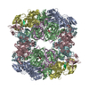
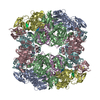
| Entry | Database: PDB / ID: 4a8d | ||||||
|---|---|---|---|---|---|---|---|
| Title | DegP dodecamer with bound OMP | ||||||
 Components Components |
| ||||||
 Keywords Keywords | HYDROLASE/TRANSPORT PROTEIN / HYDROLASE-TRANSPORT PROTEIN COMPLEX /  CHAPERONE CHAPERONE | ||||||
| Function / homology |  Function and homology informationpeptidase Do / response to temperature stimulus / porin activity / protein quality control for misfolded or incompletely synthesized proteins / pore complex / chaperone-mediated protein folding / monoatomic ion transmembrane transport / serine-type peptidase activity / cell outer membrane / protein folding ...peptidase Do / response to temperature stimulus / porin activity / protein quality control for misfolded or incompletely synthesized proteins / pore complex / chaperone-mediated protein folding / monoatomic ion transmembrane transport / serine-type peptidase activity / cell outer membrane / protein folding / virus receptor activity / outer membrane-bounded periplasmic space / peptidase activity / response to heat / response to oxidative stress / receptor-mediated virion attachment to host cell / periplasmic space / serine-type endopeptidase activity / DNA damage response / proteolysis / identical protein binding / metal ion binding / plasma membrane Function and homology informationpeptidase Do / response to temperature stimulus / porin activity / protein quality control for misfolded or incompletely synthesized proteins / pore complex / chaperone-mediated protein folding / monoatomic ion transmembrane transport / serine-type peptidase activity / cell outer membrane / protein folding ...peptidase Do / response to temperature stimulus / porin activity / protein quality control for misfolded or incompletely synthesized proteins / pore complex / chaperone-mediated protein folding / monoatomic ion transmembrane transport / serine-type peptidase activity / cell outer membrane / protein folding / virus receptor activity / outer membrane-bounded periplasmic space / peptidase activity / response to heat / response to oxidative stress / receptor-mediated virion attachment to host cell / periplasmic space / serine-type endopeptidase activity / DNA damage response / proteolysis / identical protein binding / metal ion binding / plasma membraneSimilarity search - Function | ||||||
| Biological species |  ESCHERICHIA COLI (E. coli) ESCHERICHIA COLI (E. coli) | ||||||
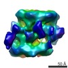
| Method | ELECTRON MICROSCOPY / single particle reconstruction / cryo EM / Resolution: 28 Å | ||||||
| Model type details | CA ATOMS ONLY, CHAIN A, B, C, D, E, F, G, H, I, J, K, L, M | ||||||
 Authors Authors | Malet, H. / Krojer, T. / Sawa, J. / Schafer, E. / Saibil, H.R. / Ehrmann, M. / Clausen, T. | ||||||
 Citation Citation | Journal: Nat Struct Mol Biol / Year: 2012 Title: Newly folded substrates inside the molecular cage of the HtrA chaperone DegQ. Authors: Hélène Malet / Flavia Canellas / Justyna Sawa / Jun Yan / Konstantinos Thalassinos / Michael Ehrmann / Tim Clausen / Helen R Saibil /  Abstract: The HtrA protein family combines chaperone and protease activities and is essential for protein quality control in many organisms. Whereas the mechanisms underlying the proteolytic function of HtrA ...The HtrA protein family combines chaperone and protease activities and is essential for protein quality control in many organisms. Whereas the mechanisms underlying the proteolytic function of HtrA proteins are well characterized, their chaperone activity remains poorly understood. Here we describe cryo-EM structures of Escherichia coli DegQ in its 12- and 24-mer states in complex with model substrates, providing a structural model of HtrA chaperone action. Up to six lysozyme substrates bind inside the DegQ 12-mer cage and are visualized in a close-to-native state. An asymmetric reconstruction reveals the binding of a well-ordered lysozyme to four DegQ protomers. DegQ PDZ domains are located adjacent to substrate density and their presence is required for chaperone activity. The substrate-interacting regions appear conserved in 12- and 24-mer cages, suggesting a common mechanism of chaperone function. #1: Journal: Nature / Year: 2008Title: Structural basis for the regulated protease and chaperone function of DegP. Authors: Tobias Krojer / Justyna Sawa / Eva Schäfer / Helen R Saibil / Michael Ehrmann / Tim Clausen /  Abstract: All organisms have to monitor the folding state of cellular proteins precisely. The heat-shock protein DegP is a protein quality control factor in the bacterial envelope that is involved in ...All organisms have to monitor the folding state of cellular proteins precisely. The heat-shock protein DegP is a protein quality control factor in the bacterial envelope that is involved in eliminating misfolded proteins and in the biogenesis of outer-membrane proteins. Here we describe the molecular mechanisms underlying the regulated protease and chaperone function of DegP from Escherichia coli. We show that binding of misfolded proteins transforms hexameric DegP into large, catalytically active 12-meric and 24-meric multimers. A structural analysis of these particles revealed that DegP represents a protein packaging device whose central compartment is adaptable to the size and concentration of substrate. Moreover, the inner cavity serves antagonistic functions. Whereas the encapsulation of folded protomers of outer-membrane proteins is protective and might allow safe transit through the periplasm, misfolded proteins are eliminated in the molecular reaction chamber. Oligomer reassembly and concomitant activation on substrate binding may also be critical in regulating other HtrA proteases implicated in protein-folding diseases. | ||||||
| History |
|
- Structure visualization
| Movie |
Movie viewer |
|---|---|
| Structure viewer | Molecule: MolmilJmol/JSmol |
- Downloads & links
-Download
| PDBx/mmCIF format | 4a8d.cif.gz | 162.6 KB | Display | PDBx/mmCIF format |
|---|---|---|---|---|
| PDB format | pdb4a8d.ent.gz | 114.9 KB | Display | PDB format |
| PDBx/mmJSON format | 4a8d.json.gz | Tree view | PDBx/mmJSON format | |
| Others |  Other downloads Other downloads |
-Validation report
| Arichive directory | https://data.pdbj.org/pub/pdb/validation_reports/a8/4a8dftp://data.pdbj.org/pub/pdb/validation_reports/a8/4a8d | HTTPS FTP |
|---|
-Related structure data
| Related structure data |  1505M 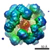 1981C 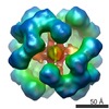 1982C 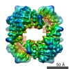 1983C 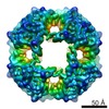 1984C 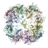 4a8aC 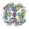 4a8bC 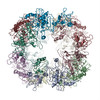 4a8cC  4a9gC M: map data used to model this data C: citing same article ( |
|---|---|
| Similar structure data |


-Links
 PDBj
PDBj
- Assembly
| Deposited unit | 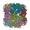
|
|---|---|
| 1 |
|
-Components
| #1: Protein | Mass: 46852.926 Da / Num. of mol.: 12 / Fragment: DEGP / Mutation: YES Source method: isolated from a genetically manipulated source Source: (gene. exp.) ESCHERICHIA COLI (E. coli) / Production host: ESCHERICHIA COLI (E. coli) / Strain (production host): CLC198 / References: UniProt: P0C0V0, peptidase Do#2: Protein | | Mass: 38336.242 Da / Num. of mol.: 1 / Source method: isolated from a natural source / Source: (natural) ESCHERICHIA COLI (E. coli) / Strain: CLC198 / References: UniProt: P06996Compound details | ENGINEERED RESIDUE IN CHAIN A, SER 236 TO ALA ENGINEERED RESIDUE IN CHAIN B, SER 236 TO ALA ...ENGINEERED | |
|---|
-Experimental details
-Experiment
| Experiment | Method: ELECTRON MICROSCOPY |
|---|---|
| EM experiment | Aggregation state: PARTICLE / 3D reconstruction method: single particle reconstruction |
- Sample preparation
| Component | Name: DEGP DODECAMER BOUND TO OMP / Type: COMPLEX |
|---|---|
| Buffer solution | Name: 300MM NACL, 50MM HEPES- NAOH / pH: 8 / Details: 300MM NACL, 50MM HEPES- NAOH |
| Specimen | Conc.: 0.16 mg/ml / Embedding applied: NO / Shadowing applied: NO / Staining applied: NO / Vitrification applied: YES |
| Specimen support | Details: OTHER |
| Vitrification | Instrument: FEI VITROBOT MARK I / Cryogen name: ETHANE Details: EMBEDDED IN VITREOUS ICE USING C-FLAT HOLEY CARBON GRIDS AND A VITROBOT AT 20C. |
- Electron microscopy imaging
| Experimental equipment |  Model: Tecnai F20 / Image courtesy: FEI Company |
|---|---|
| Microscopy | Model: FEI TECNAI F20 |
| Electron gun | Electron source: FIELD EMISSION GUN / Accelerating voltage: 200 kV / Illumination mode: FLOOD BEAM |
| Electron lens | Mode: BRIGHT FIELDBright-field microscopy / Nominal magnification: 68100 X / Calibrated magnification: 68100 X / Nominal defocus max: 2500 nm / Nominal defocus min: 1500 nm / Cs: 2 mm |
| Specimen holder | Temperature: 91 K / Tilt angle max: 0 ° / Tilt angle min: -0.5 ° |
| Image recording | Electron dose: 15 e/Å2 / Film or detector model: GENERIC CCD |
| Radiation wavelength | Relative weight: 1 |
- Processing
| EM software |
| ||||||||||||
|---|---|---|---|---|---|---|---|---|---|---|---|---|---|
| CTF correction | Details: PHASE FLIPPING | ||||||||||||
| Symmetry | Point symmetry: D3 (2x3 fold dihedral) | ||||||||||||
| 3D reconstruction | Resolution: 28 Å / Num. of particles: 6285 / Nominal pixel size: 4.44 Å / Actual pixel size: 4.44 Å Details: SUBMISSION BASED ON EXPERIMENTAL DATA FROM EMDB EMD-1505(DEPOSITION ID: 6111). Symmetry type: POINT | ||||||||||||
| Atomic model building | Protocol: RIGID BODY FIT / Space: REAL / Target criteria: Cross-correlation coefficient Details: METHOD--RIGID BODY FITTING REFINEMENT PROTOCOL--X-RAY | ||||||||||||
| Atomic model building | PDB-ID: 3CS0 | ||||||||||||
| Refinement | Highest resolution: 28 Å | ||||||||||||
| Refinement step | Cycle: LAST / Highest resolution: 28 Å
|