 Movie
Movie Controller

[English] 日本語
 Yorodumi
- PDB-3zif: Cryo-EM structures of two intermediates provide insight into aden... -
+ Open data

- Basic information
| Entry | Database: PDB / ID: 3zif | ||||||
|---|---|---|---|---|---|---|---|
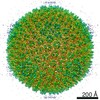
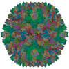
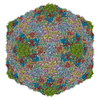
| Title | Cryo-EM structures of two intermediates provide insight into adenovirus assembly and disassembly | ||||||
 Components Components |
| ||||||
 Keywords Keywords |  VIRUS / ASSEMBLY INTERMEDIATE VIRUS / ASSEMBLY INTERMEDIATE | ||||||
| Function / homology |  Function and homology information Function and homology informationhexon binding / viral capsid, decoration / T=25 icosahedral viral capsid / microtubule-dependent intracellular transport of viral material towards nucleus / viral process / endocytosis involved in viral entry into host cell / viral capsid / symbiont entry into host cell / host cell nucleus / virion attachment to host cell / structural molecule activitySimilarity search - Function | ||||||
| Biological species |  BOVINE ADENOVIRUS 3 BOVINE ADENOVIRUS 3 | ||||||
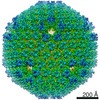
| Method | ELECTRON MICROSCOPY / single particle reconstruction / cryo EM / Resolution: 4.5 Å | ||||||
 Authors Authors | Cheng, L. / Huang, X. / Li, X. / Xiong, W. / Sun, W. / Yang, C. / Zhang, K. / Wang, Y. / Liu, H. / Ji, G. ...Cheng, L. / Huang, X. / Li, X. / Xiong, W. / Sun, W. / Yang, C. / Zhang, K. / Wang, Y. / Liu, H. / Ji, G. / Sun, F. / Zheng, C. / Zhu, P. | ||||||
 Citation Citation | Journal: Virology / Year: 2014 Title: Cryo-EM structures of two bovine adenovirus type 3 intermediates. Authors: Lingpeng Cheng / Xiaoxing Huang / Xiaomin Li / Wei Xiong / Wei Sun / Chongwen Yang / Kai Zhang / Ying Wang / Hongrong Liu / Xiaojun Huang / Gang Ji / Fei Sun / Congyi Zheng / Ping Zhu /  Abstract: Adenoviruses (Ads) infect hosts from all vertebrate species and have been investigated as vaccine vectors. We report here near-atomic structures of two bovine Ad type 3 (BAd3) intermediates obtained ...Adenoviruses (Ads) infect hosts from all vertebrate species and have been investigated as vaccine vectors. We report here near-atomic structures of two bovine Ad type 3 (BAd3) intermediates obtained by cryo-electron microscopy. A comparison between the two intermediate structures reveals that the differences are localized in the fivefold vertex region, while their facet structures are identical. The overall facet structure of BAd3 exhibits a similar structure to human Ads; however, BAd3 protein IX has a unique conformation. Mass spectrometry and cryo-electron tomography analyses indicate that one intermediate structure represents the stage during DNA encapsidation, whilst the other intermediate structure represents a later stage. These results also suggest that cleavage of precursor protein VI occurs during, rather than after, the DNA encapsidation process. Overall, our results provide insights into the mechanism of Ad assembly, and allow the first structural comparison between human and nonhuman Ads at backbone level. | ||||||
| History |
| ||||||
| Remark 700 | SHEET DETERMINATION METHOD: DSSP THE SHEETS PRESENTED AS "AK" IN EACH CHAIN ON SHEET RECORDS BELOW ... SHEET DETERMINATION METHOD: DSSP THE SHEETS PRESENTED AS "AK" IN EACH CHAIN ON SHEET RECORDS BELOW IS ACTUALLY AN 6-STRANDED BARREL THIS IS REPRESENTED BY A 7-STRANDED SHEET IN WHICH THE FIRST AND LAST STRANDS ARE IDENTICAL. THE SHEETS PRESENTED AS "DK" IN EACH CHAIN ON SHEET RECORDS BELOW IS ACTUALLY AN 6-STRANDED BARREL THIS IS REPRESENTED BY A 7-STRANDED SHEET IN WHICH THE FIRST AND LAST STRANDS ARE IDENTICAL. THE SHEETS PRESENTED AS "GK" IN EACH CHAIN ON SHEET RECORDS BELOW IS ACTUALLY AN 6-STRANDED BARREL THIS IS REPRESENTED BY A 7-STRANDED SHEET IN WHICH THE FIRST AND LAST STRANDS ARE IDENTICAL. THE SHEETS PRESENTED AS "JK" IN EACH CHAIN ON SHEET RECORDS BELOW IS ACTUALLY AN 6-STRANDED BARREL THIS IS REPRESENTED BY A 7-STRANDED SHEET IN WHICH THE FIRST AND LAST STRANDS ARE IDENTICAL. |
- Structure visualization
| Movie |
Movie viewer |
|---|---|
| Structure viewer | Molecule: MolmilJmol/JSmol |
- Downloads & links
-Download
| PDBx/mmCIF format | 3zif.cif.gz | 2 MB | Display | PDBx/mmCIF format |
|---|---|---|---|---|
| PDB format | pdb3zif.ent.gz | 1.7 MB | Display | PDB format |
| PDBx/mmJSON format | 3zif.json.gz | Tree view | PDBx/mmJSON format | |
| Others |  Other downloads Other downloads |
-Validation report
| Arichive directory | https://data.pdbj.org/pub/pdb/validation_reports/zi/3zifftp://data.pdbj.org/pub/pdb/validation_reports/zi/3zif | HTTPS FTP |
|---|
-Related structure data
| Related structure data |  2273MC  2272C M: map data used to model this data C: citing same article ( |
|---|---|
| Similar structure data |


-Links
 PDBj
PDBj


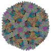
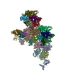
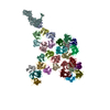
- Assembly
| Deposited unit | 
|
|---|---|
| 1 | x 60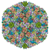
|
| 2 |
|
| 3 | x 5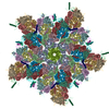
|
| 4 | x 6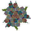
|
| 5 | 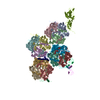
|
| Symmetry | Point symmetry: (Schoenflies symbol: I (icosahedral)) |
| Noncrystallographic symmetry (NCS) | NCS oper: (Code: given / Matrix: (1), |
-Components
| #1: Protein | / LATE PROTEIN 2 / BOVINE ADENOVIRUS 3 Mass: 103148.008 Da / Num. of mol.: 12 / Source method: isolated from a natural source / Source: (natural) BOVINE ADENOVIRUS 3 / References: UniProt: P03278#2: Protein | | / BOVINE ADENOVIRUS 3Mass: 55135.320 Da / Num. of mol.: 1 / Source method: isolated from a natural source / Source: (natural) BOVINE ADENOVIRUS 3 / References: UniProt: O71096#3: Protein | Mass: 13719.456 Da / Num. of mol.: 4 / Source method: isolated from a natural source / Source: (natural) BOVINE ADENOVIRUS 3 / References: UniProt: Q64845#4: Protein | | Mass: 23746.486 Da / Num. of mol.: 1 / Source method: isolated from a natural source / Source: (natural) BOVINE ADENOVIRUS 3 / References: UniProt: O92788 |
|---|
-Experimental details
-Experiment
| Experiment | Method: ELECTRON MICROSCOPY |
|---|---|
| EM experiment | Aggregation state: PARTICLE / 3D reconstruction method: single particle reconstruction |
- Sample preparation
| Component | Name: BOVINE ADENOVIRUS TYPE 3 / Type: VIRUS |
|---|---|
| Buffer solution | Name: PBS / Details: PBS |
| Specimen | Embedding applied: NO / Shadowing applied: NO / Staining applied: NO / Vitrification applied: YES |
| Specimen support | Details: HOLEY CARBON |
| Vitrification | Instrument: FEI VITROBOT MARK II / Cryogen name: ETHANE / Details: LIQUID ETHAN |
- Electron microscopy imaging
| Experimental equipment |  Model: Titan Krios / Image courtesy: FEI Company |
|---|---|
| Microscopy | Model: FEI TITAN KRIOS / Date: Feb 2, 2011 |
| Electron gun | Electron source: TUNGSTEN HAIRPIN / Accelerating voltage: 300 kV / Illumination mode: FLOOD BEAM |
| Electron lens | Mode: BRIGHT FIELDBright-field microscopy / Nominal magnification: 75000 X / Calibrated magnification: 125390 X / Nominal defocus max: 3500 nm / Nominal defocus min: 1000 nm / Cs: 2.7 mm |
| Image recording | Electron dose: 25 e/Å2 / Film or detector model: GENERIC GATAN |
| Image scans | Num. digital images: 1275 |
| Radiation wavelength | Relative weight: 1 |
- Processing
| EM software |
| ||||||||||||
|---|---|---|---|---|---|---|---|---|---|---|---|---|---|
| CTF correction | Details: INDIVIDUAL IMAGES | ||||||||||||
| Symmetry | Point symmetry: I (icosahedral) | ||||||||||||
| 3D reconstruction | Method: CROSS-COMMON LINES / Resolution: 4.5 Å / Num. of particles: 11910 / Actual pixel size: 1.16 Å Details: SUBMISSION BASED ON EXPERIMENTAL DATA FROM EMDB EMD-2273. (DEPOSITION ID: 11337). Symmetry type: POINT | ||||||||||||
| Refinement | Highest resolution: 4.5 Å | ||||||||||||
| Refinement step | Cycle: LAST / Highest resolution: 4.5 Å
|
