 Movie
Movie Controller


+ Open data

- Basic information
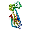
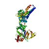
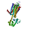
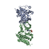
| Entry | Database: PDB / ID: 3v2w | ||||||
|---|---|---|---|---|---|---|---|
| Title | Crystal Structure of a Lipid G protein-Coupled Receptor at 3.35A | ||||||
 Components Components | Sphingosine 1-phosphate receptor 1, Lysozyme chimera | ||||||
 Keywords Keywords |  HYDROLASE / sphingosine / EDG receptor / lipid receptor / multiple sclerosis / autoimmunity / Structural Genomics / PSI-Biology / GPCR Network / GPCR / membrane protein / G protein coupled receptor / membrane HYDROLASE / sphingosine / EDG receptor / lipid receptor / multiple sclerosis / autoimmunity / Structural Genomics / PSI-Biology / GPCR Network / GPCR / membrane protein / G protein coupled receptor / membrane | ||||||
| Function / homology |  Function and homology information Function and homology informationcardiac muscle tissue growth involved in heart morphogenesis / sphingosine-1-phosphate receptor activity / sphingolipid binding / blood vessel maturation / Lysosphingolipid and LPA receptors / T cell migration / endothelial cell differentiation / heart trabecula morphogenesis / regulation of metabolic process / regulation of bone mineralization ...cardiac muscle tissue growth involved in heart morphogenesis / sphingosine-1-phosphate receptor activity / sphingolipid binding / blood vessel maturation / Lysosphingolipid and LPA receptors / T cell migration / endothelial cell differentiation / heart trabecula morphogenesis / regulation of metabolic process / regulation of bone mineralization / sphingosine-1-phosphate receptor signaling pathway / leukocyte chemotaxis / regulation of bone resorption / positive regulation of positive chemotaxis / lamellipodium assembly / negative regulation of stress fiber assembly / transmission of nerve impulse / regulation of cell adhesion / viral release from host cell by cytolysis / adenylate cyclase-inhibiting G protein-coupled receptor signaling pathway / peptidoglycan catabolic process / G protein-coupled receptor binding / G protein-coupled receptor activity / positive regulation of smooth muscle cell proliferation / brain development / adenylate cyclase-activating G protein-coupled receptor signaling pathway / neuron differentiation / chemotaxis / cell migration / cell wall macromolecule catabolic process / lysozyme / lysozyme activity / phospholipase C-activating G protein-coupled receptor signaling pathway / actin cytoskeleton organization / angiogenesis / Interleukin-4 and Interleukin-13 signaling / cell population proliferation / host cell cytoplasm / Potential therapeutics for SARS / cell adhesion / endosome / positive regulation of cell migration / defense response to bacterium / membrane raft / G protein-coupled receptor signaling pathway / external side of plasma membrane / intracellular membrane-bounded organelle / positive regulation of transcription by RNA polymerase II / nucleoplasm / plasma membrane / cytoplasmSimilarity search - Function | ||||||
| Biological species |  Homo sapiens (human) Homo sapiens (human) Enterobacteria phage T4 (virus) Enterobacteria phage T4 (virus) | ||||||
| Method | X-RAY DIFFRACTION / SYNCHROTRON / MOLECULAR REPLACEMENT / Resolution: 3.35 Å | ||||||
 Authors Authors | Hanson, M.A. / Roth, C.B. / Jo, E. / Griffith, M.T. / Scott, F.L. / Reinhart, G. / Desale, H. / Clemons, B. / Cahalan, S.M. / Schuerer, S.C. ...Hanson, M.A. / Roth, C.B. / Jo, E. / Griffith, M.T. / Scott, F.L. / Reinhart, G. / Desale, H. / Clemons, B. / Cahalan, S.M. / Schuerer, S.C. / Sanna, M.G. / Han, G.W. / Kuhn, P. / Rosen, H. / Stevens, R.C. / GPCR Network (GPCR) | ||||||
 Citation Citation | Journal: Science / Year: 2012 Title: Crystal structure of a lipid G protein-coupled receptor. Authors: Hanson, M.A. / Roth, C.B. / Jo, E. / Griffith, M.T. / Scott, F.L. / Reinhart, G. / Desale, H. / Clemons, B. / Cahalan, S.M. / Schuerer, S.C. / Sanna, M.G. / Han, G.W. / Kuhn, P. / Rosen, H. / Stevens, R.C. | ||||||
| History |
|
- Structure visualization
| Structure viewer | Molecule: MolmilJmol/JSmol |
|---|
- Downloads & links
-Download
| PDBx/mmCIF format | 3v2w.cif.gz | 188.1 KB | Display | PDBx/mmCIF format |
|---|---|---|---|---|
| PDB format | pdb3v2w.ent.gz | 148.1 KB | Display | PDB format |
| PDBx/mmJSON format | 3v2w.json.gz | Tree view | PDBx/mmJSON format | |
| Others |  Other downloads Other downloads |
-Validation report
| Arichive directory | https://data.pdbj.org/pub/pdb/validation_reports/v2/3v2wftp://data.pdbj.org/pub/pdb/validation_reports/v2/3v2w | HTTPS FTP |
|---|
-Related structure data


-Links
 PDBj
PDBj


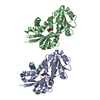
- Assembly
| Deposited unit | 
| ||||||||
|---|---|---|---|---|---|---|---|---|---|
| 1 |
| ||||||||
| Unit cell |
| ||||||||
| Details | THE AUTHOR STATES THAT THE BIOLOGICAL UNIT OF THIS PROTEIN IS UNKNOWN. |
-Components
| #1: Protein | Mass: 58973.168 Da / Num. of mol.: 1 / Mutation: C1054T, C1097A Source method: isolated from a genetically manipulated source Source: (gene. exp.) Homo sapiens (human), (gene. exp.) Enterobacteria phage T4 (virus)Gene: S1PR1, CHEDG1, EDG1 / Plasmid: pFASTBAC / Production host:   Spodoptera frugiperda (fall armyworm) / References: UniProt: P21453, UniProt: P00720, lysozyme Spodoptera frugiperda (fall armyworm) / References: UniProt: P21453, UniProt: P00720, lysozyme |
|---|---|
| #2: Chemical | ChemComp-ML5 / {(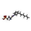  Mass: 342.370 Da / Num. of mol.: 1 / Source method: obtained synthetically / Formula: C16H27N2O4P Mass: 342.370 Da / Num. of mol.: 1 / Source method: obtained synthetically / Formula: C16H27N2O4P |
| #3: Sugar | ChemComp-NAG / N-Acetylglucosamine  Type: D-saccharide, beta linking / Mass: 221.208 Da / Num. of mol.: 1 Type: D-saccharide, beta linking / Mass: 221.208 Da / Num. of mol.: 1Source method: isolated from a genetically manipulated source Formula: C8H15NO6 |
| Sequence details | THE AUTHORS STATE THAT THE SEQUENCE "NV" IS THE ORIGINAL SEQUENCE FROM ENDOTHELIUM, AS OPPOSED TO ...THE AUTHORS STATE THAT THE SEQUENCE "NV" IS THE ORIGINAL SEQUENCE FROM ENDOTHELIU |
-Experimental details
-Experiment
| Experiment | Method: X-RAY DIFFRACTION / Number of used crystals: 33 |
|---|
- Sample preparation
| Crystal | Density Matthews: 2.61 Å3/Da / Density % sol: 52.93 % |
|---|---|
| Crystal grow | Temperature: 287 K / Method: lupuc cubic phase Details: 0.1M Tricine, 34-36% PEG400, 80mM sodium citrate and 4% glycerol, Lupuc cubic phase, temperature 287K |
-Data collection
| Diffraction |
| ||||||||||||
|---|---|---|---|---|---|---|---|---|---|---|---|---|---|
| Diffraction source |
| ||||||||||||
| Detector | Type: MARMOSAIC 300 mm CCD / Detector: CCD / Date: Jan 1, 2009 | ||||||||||||
| Radiation | Protocol: SINGLE WAVELENGTH / Monochromatic (M) / Laue (L): M / Scattering type: x-ray | ||||||||||||
| Radiation wavelength | Relative weight: 1 | ||||||||||||
| Reflection | Resolution: 3.35→20 Å / Num. obs: 8293 / % possible obs: 90.1 % / Redundancy: 2.7 % / Biso Wilson estimate: 101.89 Å2 / Rmerge(I) obs: 0.18 / Net I/σ(I): 3.5 | ||||||||||||
| Reflection shell | Resolution: 3.35→3.53 Å / Redundancy: 2 % / Rmerge(I) obs: 0.78 / Mean I/σ(I) obs: 1.1 / % possible all: 80.8 |

- Processing
| Software |
| ||||||||||||||||||||||||||||||||||||||||||||||||||||||||||||||||||||||||||||||||||||||||||||||||||||||||||||||||||
|---|---|---|---|---|---|---|---|---|---|---|---|---|---|---|---|---|---|---|---|---|---|---|---|---|---|---|---|---|---|---|---|---|---|---|---|---|---|---|---|---|---|---|---|---|---|---|---|---|---|---|---|---|---|---|---|---|---|---|---|---|---|---|---|---|---|---|---|---|---|---|---|---|---|---|---|---|---|---|---|---|---|---|---|---|---|---|---|---|---|---|---|---|---|---|---|---|---|---|---|---|---|---|---|---|---|---|---|---|---|---|---|---|---|---|---|
| Refinement | Method to determine structure: MOLECULAR REPLACEMENT Starting model: 7TM of b2AR and T4L Resolution: 3.35→19.65 Å / Cor.coef. Fo:Fc: 0.887 / Cor.coef. Fo:Fc free: 0.8232 / Cross valid method: THROUGHOUT / σ(F): 0
| ||||||||||||||||||||||||||||||||||||||||||||||||||||||||||||||||||||||||||||||||||||||||||||||||||||||||||||||||||
| Displacement parameters | Biso mean: 76.97 Å2
| ||||||||||||||||||||||||||||||||||||||||||||||||||||||||||||||||||||||||||||||||||||||||||||||||||||||||||||||||||
| Refine analyze | Luzzati coordinate error obs: 0.704 Å | ||||||||||||||||||||||||||||||||||||||||||||||||||||||||||||||||||||||||||||||||||||||||||||||||||||||||||||||||||
| Refinement step | Cycle: LAST / Resolution: 3.35→19.65 Å
| ||||||||||||||||||||||||||||||||||||||||||||||||||||||||||||||||||||||||||||||||||||||||||||||||||||||||||||||||||
| Refine LS restraints |
| ||||||||||||||||||||||||||||||||||||||||||||||||||||||||||||||||||||||||||||||||||||||||||||||||||||||||||||||||||
| LS refinement shell | Resolution: 3.35→3.75 Å / Total num. of bins used: 5
| ||||||||||||||||||||||||||||||||||||||||||||||||||||||||||||||||||||||||||||||||||||||||||||||||||||||||||||||||||
| Refinement TLS params. | Method: refined / Origin x: 17.812 Å / Origin y: 18.6984 Å / Origin z: 13.8746 Å
| ||||||||||||||||||||||||||||||||||||||||||||||||||||||||||||||||||||||||||||||||||||||||||||||||||||||||||||||||||
| Refinement TLS group | Selection details: chain A |
