 Movie
Movie Controller


+ Open data

- Basic information
| Entry | Database: PDB / ID: 3ug9 | ||||||
|---|---|---|---|---|---|---|---|
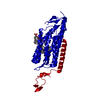
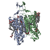
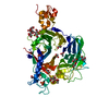
| Title | Crystal Structure of the Closed State of Channelrhodopsin | ||||||
 Components Components | Archaeal-type opsin 1, Archaeal-type opsin 2 | ||||||
 Keywords Keywords |  MEMBRANE PROTEIN / microbialrhodopsin / seven-transmembrane / light-gated cation channel MEMBRANE PROTEIN / microbialrhodopsin / seven-transmembrane / light-gated cation channel | ||||||
| Function / homology |  Function and homology information Function and homology information | ||||||
| Biological species |   Chlamydomonas reinhardtii (plant) Chlamydomonas reinhardtii (plant) | ||||||
| Method | X-RAY DIFFRACTION / SYNCHROTRON / MOLECULAR REPLACEMENT / Resolution: 2.3 Å | ||||||
 Authors Authors | Kato, H.E. / Ishitani, R. / Nureki, O. | ||||||
 Citation Citation | Journal: Nature / Year: 2012 Title: Crystal structure of the channelrhodopsin light-gated cation channel Authors: Kato, H.E. / Zhang, F. / Yizhar, O. / Ramakrishnan, C. / Nishizawa, T. / Hirata, K. / Ito, J. / Aita, Y. / Tsukazaki, T. / Hayashi, S. / Hegemann, P. / Maturana, A.D. / Ishitani, R. / Deisseroth, K. / Nureki, O. | ||||||
| History |
|
- Structure visualization
| Structure viewer | Molecule: MolmilJmol/JSmol |
|---|
- Downloads & links
-Download
| PDBx/mmCIF format | 3ug9.cif.gz | 124.3 KB | Display | PDBx/mmCIF format |
|---|---|---|---|---|
| PDB format | pdb3ug9.ent.gz | 101.1 KB | Display | PDB format |
| PDBx/mmJSON format | 3ug9.json.gz | Tree view | PDBx/mmJSON format | |
| Others |  Other downloads Other downloads |
-Validation report
| Arichive directory | https://data.pdbj.org/pub/pdb/validation_reports/ug/3ug9ftp://data.pdbj.org/pub/pdb/validation_reports/ug/3ug9 | HTTPS FTP |
|---|
-Related structure data
| Related structure data |  3uga |
|---|---|
| Similar structure data |


-Links
 PDBj
PDBj


- Assembly
| Deposited unit | 
| ||||||||
|---|---|---|---|---|---|---|---|---|---|
| 1 | 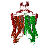
| ||||||||
| Unit cell |
|
-Components
| #1: Protein | Mass: 37441.984 Da / Num. of mol.: 1 Source method: isolated from a genetically manipulated source Details: chimera protein of Archaeal-type opsin 1 (UNP residues 24-245) and Archaeal-type opsin 2 (UNP residues 207-309) from Chlamydomonas reinhardtii Source: (gene. exp.) Chlamydomonas reinhardtii (plant) / Gene: acop1, acop2, CHLREDRAFT_182032, cop4, CSOB / Plasmid: pFastBac / Cell line (production host): Sf9 / Production host:   Spodoptera frugiperda (fall armyworm) / References: UniProt: Q93WP2, UniProt: Q8RUT8 Spodoptera frugiperda (fall armyworm) / References: UniProt: Q93WP2, UniProt: Q8RUT8 | ||
|---|---|---|---|
| #2: Chemical | ChemComp-RET / Retinal  Mass: 284.436 Da / Num. of mol.: 1 / Source method: obtained synthetically / Formula: C20H28O Mass: 284.436 Da / Num. of mol.: 1 / Source method: obtained synthetically / Formula: C20H28O | ||
| #3: Chemical | ChemComp-OLA / Oleic acid  Mass: 282.461 Da / Num. of mol.: 6 / Source method: obtained synthetically / Formula: C18H34O2 Mass: 282.461 Da / Num. of mol.: 6 / Source method: obtained synthetically / Formula: C18H34O2#4: Water | ChemComp-HOH / | Water Mass: 18.015 Da / Num. of mol.: 43 / Source method: isolated from a natural source / Formula: H2O Mass: 18.015 Da / Num. of mol.: 43 / Source method: isolated from a natural source / Formula: H2O |
-Experimental details
-Experiment
| Experiment | Method: X-RAY DIFFRACTION / Number of used crystals: 1 |
|---|
- Sample preparation
| Crystal | Density Matthews: 2.49 Å3/Da / Density % sol: 50.55 % |
|---|---|
| Crystal grow | Temperature: 293 K / Method: lipidic cubic phase / pH: 6 Details: 30% PEG 500 DME, 100mM Na Citrate (pH6.0), 100mM MgCl2, 100mM NaCl, 100mM (NH4)2SO4, lipid cubic phase, temperature 293K |
-Data collection
| Diffraction | Mean temperature: 100 K | ||||||||||||||||||||||||||||||||||||||||||||||||||||||||||||||||||||||||||||||||||||||||||||||||||||
|---|---|---|---|---|---|---|---|---|---|---|---|---|---|---|---|---|---|---|---|---|---|---|---|---|---|---|---|---|---|---|---|---|---|---|---|---|---|---|---|---|---|---|---|---|---|---|---|---|---|---|---|---|---|---|---|---|---|---|---|---|---|---|---|---|---|---|---|---|---|---|---|---|---|---|---|---|---|---|---|---|---|---|---|---|---|---|---|---|---|---|---|---|---|---|---|---|---|---|---|---|---|
| Diffraction source | Source: SYNCHROTRON / Site: SLS  / Beamline: X06SA / Wavelength: 0.9795 Å / Beamline: X06SA / Wavelength: 0.9795 Å | ||||||||||||||||||||||||||||||||||||||||||||||||||||||||||||||||||||||||||||||||||||||||||||||||||||
| Detector | Type: PSI PILATUS 6M / Detector: PIXEL / Date: Sep 2, 2011 | ||||||||||||||||||||||||||||||||||||||||||||||||||||||||||||||||||||||||||||||||||||||||||||||||||||
| Radiation | Protocol: SINGLE WAVELENGTH / Monochromatic (M) / Laue (L): M / Scattering type: x-ray | ||||||||||||||||||||||||||||||||||||||||||||||||||||||||||||||||||||||||||||||||||||||||||||||||||||
| Radiation wavelength | Wavelength: 0.9795 Å / Relative weight: 1 | ||||||||||||||||||||||||||||||||||||||||||||||||||||||||||||||||||||||||||||||||||||||||||||||||||||
| Reflection | Resolution: 2.3→37.8 Å / Num. obs: 18985 / % possible obs: 97.9 % / Observed criterion σ(I): -3 / Biso Wilson estimate: 53 Å2 / Rmerge(I) obs: 0.08 / Net I/σ(I): 16.5 | ||||||||||||||||||||||||||||||||||||||||||||||||||||||||||||||||||||||||||||||||||||||||||||||||||||
| Reflection shell | Diffraction-ID: 1
|
- Processing
| Software |
| ||||||||||||||||||||||||||||||||||||||||||||||||||||||||||||||||||||||||||||||||||||||||||||||||||||
|---|---|---|---|---|---|---|---|---|---|---|---|---|---|---|---|---|---|---|---|---|---|---|---|---|---|---|---|---|---|---|---|---|---|---|---|---|---|---|---|---|---|---|---|---|---|---|---|---|---|---|---|---|---|---|---|---|---|---|---|---|---|---|---|---|---|---|---|---|---|---|---|---|---|---|---|---|---|---|---|---|---|---|---|---|---|---|---|---|---|---|---|---|---|---|---|---|---|---|---|---|---|
| Refinement | Method to determine structure: MOLECULAR REPLACEMENT / Resolution: 2.3→36.569 Å / Occupancy max: 1 / Occupancy min: 0.5 / FOM work R set: 0.8366 / SU ML: 0.78 / σ(F): 1.34 / Phase error: 23.26 / Stereochemistry target values: ML
| ||||||||||||||||||||||||||||||||||||||||||||||||||||||||||||||||||||||||||||||||||||||||||||||||||||
| Solvent computation | Shrinkage radii: 0.73 Å / VDW probe radii: 1 Å / Solvent model: FLAT BULK SOLVENT MODEL / Bsol: 37.334 Å2 / ksol: 0.318 e/Å3 | ||||||||||||||||||||||||||||||||||||||||||||||||||||||||||||||||||||||||||||||||||||||||||||||||||||
| Displacement parameters | Biso max: 165.72 Å2 / Biso mean: 51.5985 Å2 / Biso min: 21.08 Å2
| ||||||||||||||||||||||||||||||||||||||||||||||||||||||||||||||||||||||||||||||||||||||||||||||||||||
| Refinement step | Cycle: LAST / Resolution: 2.3→36.569 Å
| ||||||||||||||||||||||||||||||||||||||||||||||||||||||||||||||||||||||||||||||||||||||||||||||||||||
| Refine LS restraints |
| ||||||||||||||||||||||||||||||||||||||||||||||||||||||||||||||||||||||||||||||||||||||||||||||||||||
| LS refinement shell | Refine-ID: X-RAY DIFFRACTION / Total num. of bins used: 12
| ||||||||||||||||||||||||||||||||||||||||||||||||||||||||||||||||||||||||||||||||||||||||||||||||||||
| Refinement TLS params. | Method: refined / Refine-ID: X-RAY DIFFRACTION
| ||||||||||||||||||||||||||||||||||||||||||||||||||||||||||||||||||||||||||||||||||||||||||||||||||||
| Refinement TLS group |
|
