 Movie
Movie Controller

[English] 日本語
 Yorodumi
- PDB-3nry: Insights into anti-parallel microtubule crosslinking by PRC1, a c... -
+ Open data

- Basic information
| Entry | Database: PDB / ID: 3nry | ||||||
|---|---|---|---|---|---|---|---|
| Title | Insights into anti-parallel microtubule crosslinking by PRC1, a conserved microtubule binding protein | ||||||
 Components Components | Protein regulator of cytokinesis 1 | ||||||
 Keywords Keywords |  PROTEIN BINDING / spectrin fold / microtubule binding protein PROTEIN BINDING / spectrin fold / microtubule binding protein | ||||||
| Function / homology |  Function and homology informationcontractile ring / mitotic spindle midzone assembly / mitotic spindle elongation / mitotic spindle midzone / RHO GTPases activate CIT / intercellular bridge / kinesin binding / regulation of cytokinesis / spindle microtubule / spindle ...contractile ring / mitotic spindle midzone assembly / mitotic spindle elongation / mitotic spindle midzone / RHO GTPases activate CIT / intercellular bridge / kinesin binding / regulation of cytokinesis / spindle microtubule / spindle / microtubule cytoskeleton organization / spindle pole / microtubule cytoskeleton / chromosome / midbody / microtubule binding / cell division / positive regulation of cell population proliferation / protein kinase binding / nucleoplasm / identical protein binding / nucleus / plasma membrane / cytosol / cytoplasm Function and homology informationcontractile ring / mitotic spindle midzone assembly / mitotic spindle elongation / mitotic spindle midzone / RHO GTPases activate CIT / intercellular bridge / kinesin binding / regulation of cytokinesis / spindle microtubule / spindle ...contractile ring / mitotic spindle midzone assembly / mitotic spindle elongation / mitotic spindle midzone / RHO GTPases activate CIT / intercellular bridge / kinesin binding / regulation of cytokinesis / spindle microtubule / spindle / microtubule cytoskeleton organization / spindle pole / microtubule cytoskeleton / chromosome / midbody / microtubule binding / cell division / positive regulation of cell population proliferation / protein kinase binding / nucleoplasm / identical protein binding / nucleus / plasma membrane / cytosol / cytoplasmSimilarity search - Function | ||||||
| Biological species |  Homo sapiens (human) Homo sapiens (human) | ||||||
| Method | X-RAY DIFFRACTION / SYNCHROTRON / SAD / Resolution: 2 Å | ||||||
 Authors Authors | Kapoor, T.M. / Subramanian, R. / Wilson-Kubalek, E.M. / Arthur, C.P. / Bick, M.J. / Campbell, E.A. / Darst, S.A. / Milligan, R.A. | ||||||
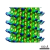
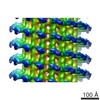
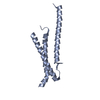
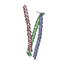
 Citation Citation | Journal: Cell / Year: 2010 Title: Insights into antiparallel microtubule crosslinking by PRC1, a conserved nonmotor microtubule binding protein. Authors: Radhika Subramanian / Elizabeth M Wilson-Kubalek / Christopher P Arthur / Matthew J Bick / Elizabeth A Campbell / Seth A Darst / Ronald A Milligan / Tarun M Kapoor /  Abstract: Formation of microtubule architectures, required for cell shape maintenance in yeast, directional cell expansion in plants and cytokinesis in eukaryotes, depends on antiparallel microtubule ...Formation of microtubule architectures, required for cell shape maintenance in yeast, directional cell expansion in plants and cytokinesis in eukaryotes, depends on antiparallel microtubule crosslinking by the conserved MAP65 protein family. Here, we combine structural and single molecule fluorescence methods to examine how PRC1, the human MAP65, crosslinks antiparallel microtubules. We find that PRC1's microtubule binding is mediated by a structured domain with a spectrin-fold and an unstructured Lys/Arg-rich domain. These two domains, at each end of a homodimer, are connected by a linkage that is flexible on single microtubules, but forms well-defined crossbridges between antiparallel filaments. Further, we show that PRC1 crosslinks are compliant and do not substantially resist filament sliding by motor proteins in vitro. Together, our data show how MAP65s, by combining structural flexibility and rigidity, tune microtubule associations to establish crosslinks that selectively "mark" antiparallel overlap in dynamic cytoskeletal networks. | ||||||
| History |
|
- Structure visualization
| Structure viewer | Molecule: MolmilJmol/JSmol |
|---|
- Downloads & links
-Download
| PDBx/mmCIF format | 3nry.cif.gz | 39.6 KB | Display | PDBx/mmCIF format |
|---|---|---|---|---|
| PDB format | pdb3nry.ent.gz | 28.2 KB | Display | PDB format |
| PDBx/mmJSON format | 3nry.json.gz | Tree view | PDBx/mmJSON format | |
| Others |  Other downloads Other downloads |
-Validation report
| Arichive directory | https://data.pdbj.org/pub/pdb/validation_reports/nr/3nryftp://data.pdbj.org/pub/pdb/validation_reports/nr/3nry | HTTPS FTP |
|---|
-Related structure data


-Links
 PDBj
PDBj

- Assembly
| Deposited unit | 
| ||||||||
|---|---|---|---|---|---|---|---|---|---|
| 1 |
| ||||||||
| Unit cell |
|
-Components
| #1: Protein | Mass: 16072.549 Da / Num. of mol.: 1 / Fragment: UNP residues 341-466 Source method: isolated from a genetically manipulated source Source: (gene. exp.) Homo sapiens (human) / Gene: PRC1 / Production host:  Escherichia coli (E. coli) / References: UniProt: O43663 Escherichia coli (E. coli) / References: UniProt: O43663 |
|---|---|
| #2: Water | ChemComp-HOH / Water Mass: 18.015 Da / Num. of mol.: 72 / Source method: isolated from a natural source / Formula: H2O Mass: 18.015 Da / Num. of mol.: 72 / Source method: isolated from a natural source / Formula: H2O |
-Experimental details
-Experiment
| Experiment | Method: X-RAY DIFFRACTION / Number of used crystals: 1 |
|---|
- Sample preparation
| Crystal | Density Matthews: 2.11 Å3/Da / Density % sol: 41.81 % |
|---|---|
| Crystal grow | Temperature: 277 K / Method: vapor diffusion, hanging drop / pH: 9.5 Details: 1:1 v/v protein (50 mg/ml in 80 mM PIPES, pH 6.8, 1 mM MgCl2, 1 mM EDTA, 150 mM KCl) and reservoir buffer (100 mM CHES pH 9.5, 30 % PEG 3000), VAPOR DIFFUSION, HANGING DROP, temperature 277K |
-Data collection
| Diffraction | Mean temperature: 173 K |
|---|---|
| Diffraction source | Source: SYNCHROTRON / Site: APS / Beamline: 24-ID-E / Wavelength: 0.97918 Å |
| Detector | Type: ADSC QUANTUM 315 / Detector: CCD / Date: Apr 21, 2009 / Details: Single crystal side bounce |
| Radiation | Monochromator: single crystal side bounce monochromator / Protocol: SINGLE WAVELENGTH / Monochromatic (M) / Laue (L): M / Scattering type: x-ray |
| Radiation wavelength | Wavelength: 0.97918 Å / Relative weight: 1 |
| Reflection | Resolution: 2→25 Å / Num. all: 17696 / Num. obs: 17342 / % possible obs: 98.3 % / Observed criterion σ(F): 0 / Observed criterion σ(I): 0 / Redundancy: 3.5 % / Rmerge(I) obs: 0.082 / Rsym value: 0.082 / Net I/σ(I): 20.1 |
| Reflection shell | Resolution: 2→2.03 Å / Redundancy: 2.8 % / Rmerge(I) obs: 0.469 / Mean I/σ(I) obs: 2 / Num. unique all: 914 / Rsym value: 0.469 / % possible all: 97.3 |
- Processing
| Software |
| ||||||||||||||||||||||||||||||||||||||||||||||||||||||||||||||||||||||||||||||||||||||||||||||||||||||||||||||||||||||||||||||||||||||||||||||||||||||||||||||||||||||||||
|---|---|---|---|---|---|---|---|---|---|---|---|---|---|---|---|---|---|---|---|---|---|---|---|---|---|---|---|---|---|---|---|---|---|---|---|---|---|---|---|---|---|---|---|---|---|---|---|---|---|---|---|---|---|---|---|---|---|---|---|---|---|---|---|---|---|---|---|---|---|---|---|---|---|---|---|---|---|---|---|---|---|---|---|---|---|---|---|---|---|---|---|---|---|---|---|---|---|---|---|---|---|---|---|---|---|---|---|---|---|---|---|---|---|---|---|---|---|---|---|---|---|---|---|---|---|---|---|---|---|---|---|---|---|---|---|---|---|---|---|---|---|---|---|---|---|---|---|---|---|---|---|---|---|---|---|---|---|---|---|---|---|---|---|---|---|---|---|---|---|---|---|
| Refinement | Method to determine structure: SAD / Resolution: 2→25 Å / Cor.coef. Fo:Fc: 0.932 / Cor.coef. Fo:Fc free: 0.902 / SU B: 5.103 / SU ML: 0.142 / Cross valid method: THROUGHOUT / ESU R Free: 0.199 Stereochemistry target values: MAXIMUM LIKELIHOOD WITH PHASES Details: HYDROGENS HAVE BEEN ADDED IN THE RIDING POSITIONS
| ||||||||||||||||||||||||||||||||||||||||||||||||||||||||||||||||||||||||||||||||||||||||||||||||||||||||||||||||||||||||||||||||||||||||||||||||||||||||||||||||||||||||||
| Solvent computation | Ion probe radii: 0.8 Å / Shrinkage radii: 0.8 Å / VDW probe radii: 1.4 Å / Solvent model: MASK | ||||||||||||||||||||||||||||||||||||||||||||||||||||||||||||||||||||||||||||||||||||||||||||||||||||||||||||||||||||||||||||||||||||||||||||||||||||||||||||||||||||||||||
| Displacement parameters | Biso mean: 24.778 Å2
| ||||||||||||||||||||||||||||||||||||||||||||||||||||||||||||||||||||||||||||||||||||||||||||||||||||||||||||||||||||||||||||||||||||||||||||||||||||||||||||||||||||||||||
| Refinement step | Cycle: LAST / Resolution: 2→25 Å
| ||||||||||||||||||||||||||||||||||||||||||||||||||||||||||||||||||||||||||||||||||||||||||||||||||||||||||||||||||||||||||||||||||||||||||||||||||||||||||||||||||||||||||
| Refine LS restraints |
| ||||||||||||||||||||||||||||||||||||||||||||||||||||||||||||||||||||||||||||||||||||||||||||||||||||||||||||||||||||||||||||||||||||||||||||||||||||||||||||||||||||||||||
| LS refinement shell | Resolution: 1.995→2.046 Å / Total num. of bins used: 20
|
