 Movie
Movie Controller


+ Open data

- Basic information
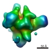
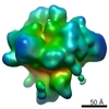
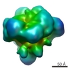
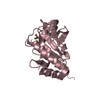
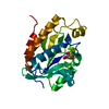
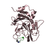
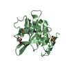
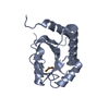
| Entry | Database: PDB / ID: 3nr5 | ||||||
|---|---|---|---|---|---|---|---|
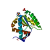
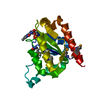
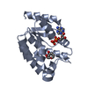
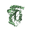
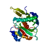
| Title | Crystal structure of human Maf1 | ||||||
 Components Components | Repressor of RNA polymerase III transcription MAF1 homolog | ||||||
 Keywords Keywords |  TRANSCRIPTION / RNA-Pol III transcriptional repressor / RNA-Pol III TRANSCRIPTION / RNA-Pol III transcriptional repressor / RNA-Pol III | ||||||
| Function / homology |  Function and homology information Function and homology informationintracellular anatomical structure / negative regulation of transcription by RNA polymerase III / RNA polymerase III core binding / RNA polymerase III type 2 promoter sequence-specific DNA binding / RNA polymerase III type 1 promoter sequence-specific DNA binding / inhibitory synapse / RNA polymerase III type 3 promoter sequence-specific DNA binding / GABA receptor binding / negative regulation of transcription by RNA polymerase I / Regulation of PTEN gene transcription ...intracellular anatomical structure / negative regulation of transcription by RNA polymerase III / RNA polymerase III core binding / RNA polymerase III type 2 promoter sequence-specific DNA binding / RNA polymerase III type 1 promoter sequence-specific DNA binding / inhibitory synapse / RNA polymerase III type 3 promoter sequence-specific DNA binding / GABA receptor binding / negative regulation of transcription by RNA polymerase I / Regulation of PTEN gene transcription / axon / intracellular membrane-bounded organelle / dendrite / nucleolus / perinuclear region of cytoplasm / nucleoplasm / nucleus / plasma membrane / cytosol / cytoplasmSimilarity search - Function | ||||||
| Biological species |  Homo sapiens (human) Homo sapiens (human) | ||||||
| Method | X-RAY DIFFRACTION / SYNCHROTRON / MAD / Resolution: 1.55 Å | ||||||
 Authors Authors | Ringel, R. / Vannini, A. / Kusser, A.G. / Berninghausen, O. / Kassavetis, G.A. / Cramer, P. | ||||||
 Citation Citation | Journal: Cell / Year: 2010 Title: Molecular basis of RNA polymerase III transcription repression by Maf1. Authors: Alessandro Vannini / Rieke Ringel / Anselm G Kusser / Otto Berninghausen / George A Kassavetis / Patrick Cramer /  Abstract: RNA polymerase III (Pol III) transcribes short RNAs required for cell growth. Under stress conditions, the conserved protein Maf1 rapidly represses Pol III transcription. We report the crystal ...RNA polymerase III (Pol III) transcribes short RNAs required for cell growth. Under stress conditions, the conserved protein Maf1 rapidly represses Pol III transcription. We report the crystal structure of Maf1 and cryo-electron microscopic structures of Pol III, an active Pol III-DNA-RNA complex, and a repressive Pol III-Maf1 complex. Binding of DNA and RNA causes ordering of the Pol III-specific subcomplex C82/34/31 that is required for transcription initiation. Maf1 binds the Pol III clamp and rearranges C82/34/31 at the rim of the active center cleft. This impairs recruitment of Pol III to a complex of promoter DNA with the initiation factors Brf1 and TBP and thus prevents closed complex formation. Maf1 does however not impair binding of a DNA-RNA scaffold and RNA synthesis. These results explain how Maf1 specifically represses transcription initiation from Pol III promoters and indicate that Maf1 also prevents reinitiation by binding Pol III during transcription elongation. | ||||||
| History |
|
- Structure visualization
| Structure viewer | Molecule: MolmilJmol/JSmol |
|---|
- Downloads & links
-Download
| PDBx/mmCIF format | 3nr5.cif.gz | 80.7 KB | Display | PDBx/mmCIF format |
|---|---|---|---|---|
| PDB format | pdb3nr5.ent.gz | 60.7 KB | Display | PDB format |
| PDBx/mmJSON format | 3nr5.json.gz | Tree view | PDBx/mmJSON format | |
| Others |  Other downloads Other downloads |
-Validation report
| Arichive directory | https://data.pdbj.org/pub/pdb/validation_reports/nr/3nr5ftp://data.pdbj.org/pub/pdb/validation_reports/nr/3nr5 | HTTPS FTP |
|---|
-Related structure data
-Links
 PDBj
PDBj- Assembly
| Deposited unit | 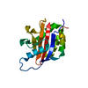
| ||||||||
|---|---|---|---|---|---|---|---|---|---|
| 1 |
| ||||||||
| Unit cell |
|
-Components
| #1: Protein | Mass: 19159.357 Da / Num. of mol.: 1 / Fragment: residues in UNP 1-35, 83-205 / Mutation: Deletion Source method: isolated from a genetically manipulated source Source: (gene. exp.) Homo sapiens (human) / Gene: MAF1 / Plasmid: pET 28b(+) / Production host:  Escherichia coli (E. coli) / References: UniProt: Q9H063 Escherichia coli (E. coli) / References: UniProt: Q9H063 |
|---|---|
| #2: Water | ChemComp-HOH / Water Mass: 18.015 Da / Num. of mol.: 141 / Source method: isolated from a natural source / Formula: H2O Mass: 18.015 Da / Num. of mol.: 141 / Source method: isolated from a natural source / Formula: H2O |
-Experimental details
-Experiment
| Experiment | Method: X-RAY DIFFRACTION / Number of used crystals: 2 |
|---|
- Sample preparation
| Crystal |
| |||||||||
|---|---|---|---|---|---|---|---|---|---|---|
| Crystal grow | Temperature: 293 K / Method: vapor diffusion, hanging drop / pH: 6 Details: 50mM MES, 175mM sodium oxalate, pH 6.0, VAPOR DIFFUSION, HANGING DROP, temperature 293K |
-Data collection
| Diffraction |
| ||||||||||||||||||
|---|---|---|---|---|---|---|---|---|---|---|---|---|---|---|---|---|---|---|---|
| Diffraction source |
| ||||||||||||||||||
| Detector |
| ||||||||||||||||||
| Radiation |
| ||||||||||||||||||
| Radiation wavelength |
| ||||||||||||||||||
| Reflection | Resolution: 1.55→26 Å / Num. all: 26137 / Num. obs: 26137 / % possible obs: 94.3 % / Observed criterion σ(F): 0 / Observed criterion σ(I): 0 |

- Processing
| Software |
| |||||||||||||||||||||||||||||||||||||||||||||||||||||||||||||||||||||||||||||
|---|---|---|---|---|---|---|---|---|---|---|---|---|---|---|---|---|---|---|---|---|---|---|---|---|---|---|---|---|---|---|---|---|---|---|---|---|---|---|---|---|---|---|---|---|---|---|---|---|---|---|---|---|---|---|---|---|---|---|---|---|---|---|---|---|---|---|---|---|---|---|---|---|---|---|---|---|---|---|
| Refinement | Method to determine structure: MAD / Resolution: 1.55→25.974 Å / Occupancy max: 1 / Occupancy min: 0.5 / FOM work R set: 0.8404 / SU ML: 0.25 / σ(F): 0 / Phase error: 22.68 / Stereochemistry target values: ML
| |||||||||||||||||||||||||||||||||||||||||||||||||||||||||||||||||||||||||||||
| Solvent computation | Shrinkage radii: 0.9 Å / VDW probe radii: 1.11 Å / Solvent model: FLAT BULK SOLVENT MODEL / Bsol: 50.129 Å2 / ksol: 0.367 e/Å3 | |||||||||||||||||||||||||||||||||||||||||||||||||||||||||||||||||||||||||||||
| Displacement parameters | Biso max: 104.56 Å2 / Biso min: 14.08 Å2
| |||||||||||||||||||||||||||||||||||||||||||||||||||||||||||||||||||||||||||||
| Refinement step | Cycle: LAST / Resolution: 1.55→25.974 Å
| |||||||||||||||||||||||||||||||||||||||||||||||||||||||||||||||||||||||||||||
| Refine LS restraints |
| |||||||||||||||||||||||||||||||||||||||||||||||||||||||||||||||||||||||||||||
| LS refinement shell | Refine-ID: X-RAY DIFFRACTION / Total num. of bins used: 10
| |||||||||||||||||||||||||||||||||||||||||||||||||||||||||||||||||||||||||||||
| Refinement TLS params. | Method: refined / Origin x: 6.4165 Å / Origin y: 13.7151 Å / Origin z: 12.0462 Å
| |||||||||||||||||||||||||||||||||||||||||||||||||||||||||||||||||||||||||||||
| Refinement TLS group | Selection details: chain A |
