 Movie
Movie Controller

[English] 日本語
 Yorodumi
- PDB-3kk5: Crystal structure of PBCV-1 VP54 fitted into a cryo-EM reconstruc... -
+ Open data

- Basic information
| Entry | Database: PDB / ID: 3kk5 | ||||||
|---|---|---|---|---|---|---|---|
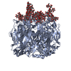
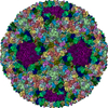
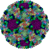
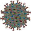
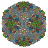
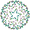
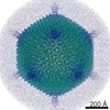
| Title | Crystal structure of PBCV-1 VP54 fitted into a cryo-EM reconstruction of the virophage Sputnik | ||||||
 Components Components | Major capsid protein | ||||||
 Keywords Keywords |  VIRUS / double jellyroll / Capsid protein / Late protein / Virion / Icosahedral virus VIRUS / double jellyroll / Capsid protein / Late protein / Virion / Icosahedral virus | ||||||
| Function / homology | Major capsid protein, N-terminal / Major capsid protein N-terminus / Major capsid protein, C-terminal / Major capsid protein, C-terminal domain superfamily / Large eukaryotic DNA virus major capsid protein / Group II dsDNA virus coat/capsid protein / viral capsid / structural molecule activity / Major capsid protein Function and homology information Function and homology information | ||||||
| Biological species |  Acanthamoeba polyphaga (eukaryote) Acanthamoeba polyphaga (eukaryote) | ||||||
| Method | ELECTRON MICROSCOPY / single particle reconstruction / cryo EM / Resolution: 10.6 Å | ||||||
 Authors Authors | Sun, S. / Rossmann, M.G. | ||||||
 Citation Citation | Journal: J Virol / Year: 2010 Title: Structural studies of the Sputnik virophage. Authors: Siyang Sun / Bernard La Scola / Valorie D Bowman / Christopher M Ryan / Julian P Whitelegge / Didier Raoult / Michael G Rossmann /  Abstract: The virophage Sputnik is a satellite virus of the giant mimivirus and is the only satellite virus reported to date whose propagation adversely affects its host virus' production. Genome sequence ...The virophage Sputnik is a satellite virus of the giant mimivirus and is the only satellite virus reported to date whose propagation adversely affects its host virus' production. Genome sequence analysis showed that Sputnik has genes related to viruses infecting all three domains of life. Here, we report structural studies of Sputnik, which show that it is about 740 A in diameter, has a T=27 icosahedral capsid, and has a lipid membrane inside the protein shell. Structural analyses suggest that the major capsid protein of Sputnik is likely to have a double jelly-roll fold, although sequence alignments do not show any detectable similarity with other viral double jelly-roll capsid proteins. Hence, the origin of Sputnik's capsid might have been derived from other viruses prior to its association with mimivirus. | ||||||
| History |
|
- Structure visualization
| Movie |
Movie viewer |
|---|---|
| Structure viewer | Molecule: MolmilJmol/JSmol |
- Downloads & links
-Download
| PDBx/mmCIF format | 3kk5.cif.gz | 168.1 KB | Display | PDBx/mmCIF format |
|---|---|---|---|---|
| PDB format | pdb3kk5.ent.gz | 117.8 KB | Display | PDB format |
| PDBx/mmJSON format | 3kk5.json.gz | Tree view | PDBx/mmJSON format | |
| Others |  Other downloads Other downloads |
-Validation report
| Arichive directory | https://data.pdbj.org/pub/pdb/validation_reports/kk/3kk5ftp://data.pdbj.org/pub/pdb/validation_reports/kk/3kk5 | HTTPS FTP |
|---|
-Related structure data
| Related structure data | 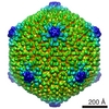 1662MC M: map data used to model this data C: citing same article ( |
|---|---|
| Similar structure data |


-Links
 PDBj
PDBj

- Assembly
| Deposited unit | 
|
|---|---|
| 1 | x 60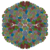
|
| 2 |
|
| 3 | x 5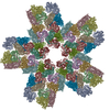
|
| 4 | x 6
|
| 5 | 
|
| Symmetry | Point symmetry: (Schoenflies symbol: I (icosahedral)) |
-Components
| #1: Protein | Mass: 48199.625 Da / Num. of mol.: 13 / Source method: isolated from a natural source / Source: (natural) Acanthamoeba polyphaga (eukaryote) / References: UniProt: P30328 |
|---|
-Experimental details
-Experiment
| Experiment | Method: ELECTRON MICROSCOPY |
|---|---|
| EM experiment | Aggregation state: PARTICLE / 3D reconstruction method: single particle reconstruction |
- Sample preparation
| Component | Name: major capsid protein of sputnik |
|---|---|
| Details of virus | Host category: PROTOZOA / Isolate: SPECIES / Type: SATELLITE |
| Natural host | Organism: Acanthamoeba polyphaga |
| Buffer solution | Name: PBS / pH: 7.4 / Details: PBS |
| Specimen | Conc.: 1 mg/ml / Embedding applied: NO / Shadowing applied: NO / Staining applied: NO / Vitrification applied: YES |
| Vitrification | Instrument: HOMEMADE PLUNGER / Cryogen name: ETHANE / Details: flash-frozen on holey grids in liquid ethane |
- Electron microscopy imaging
| Microscopy | Model: FEI/PHILIPS CM200FEG |
|---|---|
| Electron gun | Electron source: FIELD EMISSION GUN / Accelerating voltage: 200 kV / Illumination mode: FLOOD BEAM |
| Electron lens | Mode: BRIGHT FIELDBright-field microscopy / Nominal magnification: 38000 X / Calibrated magnification: 39190 X / Nominal defocus max: 3582 nm / Nominal defocus min: 767 nm / Cs: 2 mm |
| Specimen holder | Temperature: 70 K |
| Image recording | Electron dose: 20 e/Å2 / Film or detector model: GENERIC FILM |
| Radiation | Protocol: SINGLE WAVELENGTH / Monochromatic (M) / Laue (L): M / Scattering type: x-ray |
| Radiation wavelength | Relative weight: 1 |
- Processing
| EM software |
| ||||||||||||
|---|---|---|---|---|---|---|---|---|---|---|---|---|---|
| CTF correction | Details: CTF parameters were calculated for particles in each micrograph. | ||||||||||||
| Symmetry | Point symmetry: I (icosahedral) | ||||||||||||
| 3D reconstruction | Method: projection matching / Resolution: 10.6 Å / Num. of particles: 6780 / Nominal pixel size: 1.62 Å / Actual pixel size: 1.62 Å / Details: EMAN / Symmetry type: POINT | ||||||||||||
| Atomic model building | Protocol: RIGID BODY FIT / Space: REAL / Target criteria: Sumf / Details: REFINEMENT PROTOCOL--rigid body | ||||||||||||
| Atomic model building | PDB-ID: 1M3Y 1m3y Accession code: 1M3Y / Source name: PDB / Type: experimental model | ||||||||||||
| Refinement step | Cycle: LAST
|
