 Movie
Movie Controller


+ Open data

- Basic information
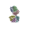
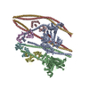
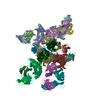
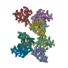
| Entry | Database: PDB / ID: 3jck | ||||||
|---|---|---|---|---|---|---|---|
| Title | Structure of the yeast 26S proteasome lid sub-complex | ||||||
 Components Components |
| ||||||
 Keywords Keywords |  HYDROLASE / Proteasome / deubiquitinase / Rpn11 / protein homeostasis HYDROLASE / Proteasome / deubiquitinase / Rpn11 / protein homeostasis | ||||||
| Function / homology |  Function and homology information Function and homology informationSAGA complex localization to transcription regulatory region / peroxisome fission / proteasome storage granule assembly / transcription export complex 2 / protein deneddylation / maintenance of DNA trinucleotide repeats / filamentous growth / COP9 signalosome / proteasome regulatory particle / proteasome regulatory particle, lid subcomplex ...SAGA complex localization to transcription regulatory region / peroxisome fission / proteasome storage granule assembly / transcription export complex 2 / protein deneddylation / maintenance of DNA trinucleotide repeats / filamentous growth / COP9 signalosome / proteasome regulatory particle / proteasome regulatory particle, lid subcomplex / mitochondrial fission / Cross-presentation of soluble exogenous antigens (endosomes) / TNFR2 non-canonical NF-kB pathway / Ub-specific processing proteases / proteasome binding / regulation of protein catabolic process / protein deubiquitination / proteasome storage granule / proteasome assembly / enzyme regulator activity / mRNA export from nucleus / protein folding chaperone / Neutrophil degranulation / proteasome complex / double-strand break repair via homologous recombination / metallopeptidase activity / ubiquitin-dependent protein catabolic process / proteasome-mediated ubiquitin-dependent protein catabolic process / ubiquitinyl hydrolase 1 / cysteine-type deubiquitinase activity / molecular adaptor activity / regulation of cell cycle / structural molecule activity / positive regulation of transcription by RNA polymerase II / mitochondrion / metal ion binding / nucleus / cytosol / cytoplasmSimilarity search - Function | ||||||
| Biological species |  Saccharomyces cerevisiae S288c (yeast) Saccharomyces cerevisiae S288c (yeast) | ||||||
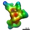
| Method | ELECTRON MICROSCOPY / single particle reconstruction / cryo EM / Resolution: 3.5 Å | ||||||
 Authors Authors | Herzik Jr., M.A. / Dambacher, C.M. / Worden, E.J. / Martin, A. / Lander, G.C. | ||||||
 Citation Citation | Journal: Elife / Year: 2016 Title: Atomic structure of the 26S proteasome lid reveals the mechanism of deubiquitinase inhibition. Authors: Corey M Dambacher / Evan J Worden / Mark A Herzik / Andreas Martin / Gabriel C Lander /  Abstract: The 26S proteasome is responsible for the selective, ATP-dependent degradation of polyubiquitinated cellular proteins. Removal of ubiquitin chains from targeted substrates at the proteasome is a ...The 26S proteasome is responsible for the selective, ATP-dependent degradation of polyubiquitinated cellular proteins. Removal of ubiquitin chains from targeted substrates at the proteasome is a prerequisite for substrate processing and is accomplished by Rpn11, a deubiquitinase within the 'lid' sub-complex. Prior to the lid's incorporation into the proteasome, Rpn11 deubiquitinase activity is inhibited to prevent unwarranted deubiquitination of polyubiquitinated proteins. Here we present the atomic model of the isolated lid sub-complex, as determined by cryo-electron microscopy at 3.5 Å resolution, revealing how Rpn11 is inhibited through its interaction with a neighboring lid subunit, Rpn5. Through mutagenesis of specific residues, we describe the network of interactions that are required to stabilize this inhibited state. These results provide significant insight into the intricate mechanisms of proteasome assembly, outlining the substantial conformational rearrangements that occur during incorporation of the lid into the 26S holoenzyme, which ultimately activates the deubiquitinase for substrate degradation. | ||||||
| History |
|
- Structure visualization
| Movie |
Movie viewer |
|---|---|
| Structure viewer | Molecule: MolmilJmol/JSmol |
- Downloads & links
-Download
| PDBx/mmCIF format | 3jck.cif.gz | 2.3 MB | Display | PDBx/mmCIF format |
|---|---|---|---|---|
| PDB format | pdb3jck.ent.gz | 2 MB | Display | PDB format |
| PDBx/mmJSON format | 3jck.json.gz | Tree view | PDBx/mmJSON format | |
| Others |  Other downloads Other downloads |
-Validation report
| Arichive directory | https://data.pdbj.org/pub/pdb/validation_reports/jc/3jckftp://data.pdbj.org/pub/pdb/validation_reports/jc/3jck | HTTPS FTP |
|---|
-Related structure data
| Related structure data | 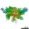 6479MC M: map data used to model this data C: citing same article ( |
|---|---|
| Similar structure data |
-Links
 PDBj
PDBj


- Assembly
| Deposited unit | 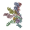
|
|---|---|
| 1 |
|
| Number of models | 5 |
-Components
-26S proteasome regulatory subunit ... , 7 types, 7 molecules ABCDEFH
| #1: Protein | Mass: 49573.766 Da / Num. of mol.: 1 Source method: isolated from a genetically manipulated source Source: (gene. exp.) Saccharomyces cerevisiae S288c (yeast) / Strain: ATCC 204508 / S288c / Cellular location: cytoplasm / Gene: RPN3, SUN2, YER021W / Plasmid: pET, pCOLA, pACYC / Production host:  Escherichia coli BL21(DE3) (bacteria) / References: UniProt: P40016 Escherichia coli BL21(DE3) (bacteria) / References: UniProt: P40016 |
|---|---|
| #2: Protein | Mass: 51840.352 Da / Num. of mol.: 1 Source method: isolated from a genetically manipulated source Source: (gene. exp.) Saccharomyces cerevisiae S288c (yeast) / Strain: ATCC 204508 / S288c / Cellular location: cytoplasm / Gene: RPN5, NAS5, YDL147W, D1572 / Plasmid: pET, pCOLA, pACYC / Production host: Escherichia coli BL21(DE3) (bacteria) / References: UniProt: Q12250 |
| #3: Protein | Mass: 49839.812 Da / Num. of mol.: 1 Source method: isolated from a genetically manipulated source Source: (gene. exp.) Saccharomyces cerevisiae S288c (yeast) / Strain: ATCC 204508 / S288c / Cellular location: cytoplasm / Gene: RPN6, NAS4, YDL097C, D2381 / Plasmid: pET, pCOLA, pACYC / Production host: Escherichia coli BL21(DE3) (bacteria) / References: UniProt: Q12377 |
| #4: Protein | Mass: 49016.367 Da / Num. of mol.: 1 Source method: isolated from a genetically manipulated source Source: (gene. exp.) Saccharomyces cerevisiae S288c (yeast) / Strain: ATCC 204508 / S288c / Cellular location: cytoplasm / Gene: RPN7, YPR108W, P8283.8 / Plasmid: pET, pCOLA, pACYC / Production host: Escherichia coli BL21(DE3) (bacteria) / References: UniProt: Q06103 |
| #5: Protein | Mass: 38365.508 Da / Num. of mol.: 1 Source method: isolated from a genetically manipulated source Source: (gene. exp.) Saccharomyces cerevisiae S288c (yeast) / Strain: ATCC 204508 / S288c / Cellular location: cytoplasm / Gene: RPN8, YOR261C, O5360 / Plasmid: pET, pCOLA, pACYC / Production host: Escherichia coli BL21(DE3) (bacteria) / References: UniProt: Q08723 |
| #6: Protein | Mass: 45839.348 Da / Num. of mol.: 1 Source method: isolated from a genetically manipulated source Source: (gene. exp.) Saccharomyces cerevisiae S288c (yeast) / Strain: ATCC 204508 / S288c / Cellular location: cytoplasm / Gene: RPN9, NAS7, YDR427W, D9461.14 / Plasmid: pET, pCOLA, pACYC / Production host: Escherichia coli BL21(DE3) (bacteria) / References: UniProt: Q04062 |
| #8: Protein | Mass: 31952.119 Da / Num. of mol.: 1 Source method: isolated from a genetically manipulated source Source: (gene. exp.) Saccharomyces cerevisiae S288c (yeast) / Strain: ATCC 204508 / S288c / Cellular location: cytoplasm / Gene: RPN12, NIN1, YFR052W / Plasmid: pET, pCOLA, pACYC / Production host: Escherichia coli BL21(DE3) (bacteria) / References: UniProt: P32496 |
-Protein , 2 types, 2 molecules GI
| #7: Protein | Mass: 34442.281 Da / Num. of mol.: 1 Source method: isolated from a genetically manipulated source Source: (gene. exp.) Saccharomyces cerevisiae S288c (yeast) / Strain: ATCC 204508 / S288c / Cellular location: cytoplasm / Gene: RPN11, MPR1, YFR004W / Plasmid: pET, pCOLA, pACYC / Production host: Escherichia coli BL21(DE3) (bacteria) / References: UniProt: P43588, ubiquitinyl hydrolase 1 |
|---|---|
| #9: Protein | Proteasome Mass: 10397.102 Da / Num. of mol.: 1 Source method: isolated from a genetically manipulated source Source: (gene. exp.) Saccharomyces cerevisiae S288c (yeast) / Strain: ATCC 204508 / S288c / Cellular location: cytoplasm / Gene: SEM1, DSH1, YDR363W-A / Plasmid: pET, pCOLA, pACYC / Production host: Escherichia coli BL21(DE3) (bacteria) / References: UniProt: O94742 |
-Non-polymers , 2 types, 2 molecules 

| #10: Chemical | ChemComp-ZN /  Mass: 65.409 Da / Num. of mol.: 1 / Source method: obtained synthetically / Formula: Zn Mass: 65.409 Da / Num. of mol.: 1 / Source method: obtained synthetically / Formula: Zn |
|---|---|
| #11: Water | ChemComp-HOH / WaterMass: 18.015 Da / Num. of mol.: 1 / Source method: isolated from a natural source / Formula: H2O |
-Experimental details
-Experiment
| Experiment | Method: ELECTRON MICROSCOPY |
|---|---|
| EM experiment | Aggregation state: PARTICLE / 3D reconstruction method: single particle reconstruction |
- Sample preparation
| Component |
| |||||||||||||||
|---|---|---|---|---|---|---|---|---|---|---|---|---|---|---|---|---|
| Molecular weight | Value: 0.37 MDa / Experimental value: YES | |||||||||||||||
| Buffer solution | Name: 50 mM HEPES, 100 mM NaCl, 100 mM KCl, 1 mM TCEP / pH: 7.5 / Details: 50 mM HEPES, 100 mM NaCl, 100 mM KCl, 1 mM TCEP | |||||||||||||||
| Specimen | Conc.: 2.5 mg/ml / Embedding applied: NO / Shadowing applied: NO / Staining applied: NO / Vitrification applied: YES | |||||||||||||||
| Specimen support | Details: Sample was applied directly to plasma-cleaned holey carbon C-flat grids (400 mesh, 1.2 micrometer holes). | |||||||||||||||
| Vitrification | Instrument: HOMEMADE PLUNGER / Cryogen name: ETHANE / Temp: 85 K / Humidity: 88 % Details: 4 uL sample was applied to the grid, blotted for 2 seconds at 4 degrees C, and plunged into liquid ethane using a manual plunger. Method: 4 uL sample was applied to the grid, blotted for 2 seconds, and plunged into liquid ethane. |
- Electron microscopy imaging
| Experimental equipment |  Model: Titan Krios / Image courtesy: FEI Company |
|---|---|
| Microscopy | Model: FEI TITAN KRIOS / Date: Feb 10, 2015 Details: Micrograph was collected in super-resolution mode with a total frame count of 38 and total exposure time of 7.6 seconds. |
| Electron gun | Electron source: FIELD EMISSION GUN / Accelerating voltage: 300 kV / Illumination mode: FLOOD BEAM |
| Electron lens | Mode: BRIGHT FIELDBright-field microscopy / Nominal magnification: 22500 X / Calibrated magnification: 38168 X / Nominal defocus max: 3200 nm / Nominal defocus min: 1600 nm / Cs: 2.7 mm Astigmatism : Objective lens astigmatism was corrected at a nominal magnification of 22,500. |
| Specimen holder | Specimen holder model: FEI TITAN KRIOS AUTOGRID HOLDER / Temperature: 87.5 K / Temperature (max): 90 K / Temperature (min): 85 K |
| Image recording | Electron dose: 43.8 e/Å2 / Film or detector model: GATAN K2 (4k x 4k) |
| Image scans | Num. digital images: 3432 |
- Processing
| EM software |
| ||||||||||||||||||||
|---|---|---|---|---|---|---|---|---|---|---|---|---|---|---|---|---|---|---|---|---|---|
| CTF correction | Details: whole micrograph | ||||||||||||||||||||
| Symmetry | Point symmetry: C1 (asymmetric) | ||||||||||||||||||||
| 3D reconstruction | Method: projection matching / Resolution: 3.5 Å / Resolution method: FSC 0.143 CUT-OFF / Num. of particles: 109396 / Nominal pixel size: 1.31 Å / Actual pixel size: 1.31 Å Details: 3D classification was performed to identify the best 109,396 particles from an initial dataset of 254,112. An ensemble of five models is provided. Each model is an approximately equivalent ...Details: 3D classification was performed to identify the best 109,396 particles from an initial dataset of 254,112. An ensemble of five models is provided. Each model is an approximately equivalent representation of the structure. (Single particle details: Image pre-processing was performed using Appion. 3D classification and reconstruction was performed with RELION.) (Single particle--Applied symmetry: C1) Symmetry type: POINT | ||||||||||||||||||||
| Refinement step | Cycle: LAST
|
