 Movie
Movie Controller


+ Open data

- Basic information
| Entry | Database: PDB / ID: 3j9i | ||||||
|---|---|---|---|---|---|---|---|
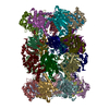
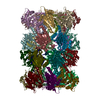
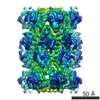
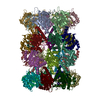
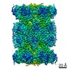
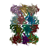
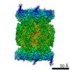
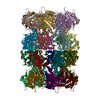
| Title | Thermoplasma acidophilum 20S proteasome | ||||||
 Components Components |
| ||||||
 Keywords Keywords |  HYDROLASE / proteasome HYDROLASE / proteasome | ||||||
| Function / homology |  Function and homology informationproteasome endopeptidase complex / proteasome core complex, beta-subunit complex / proteasome core complex, alpha-subunit complex / threonine-type endopeptidase activity / proteasomal protein catabolic process / ubiquitin-dependent protein catabolic process / endopeptidase activity / cytoplasm Function and homology informationproteasome endopeptidase complex / proteasome core complex, beta-subunit complex / proteasome core complex, alpha-subunit complex / threonine-type endopeptidase activity / proteasomal protein catabolic process / ubiquitin-dependent protein catabolic process / endopeptidase activity / cytoplasmSimilarity search - Function | ||||||
| Biological species |   Thermoplasma acidophilum (acidophilic) Thermoplasma acidophilum (acidophilic) | ||||||
| Method | ELECTRON MICROSCOPY / single particle reconstruction / cryo EM / Resolution: 3.3 Å | ||||||
 Authors Authors | Li, X. / Mooney, P. / Zheng, S. / Booth, C. / Braunfeld, M.B. / Gubbens, S. / Agard, D.A. / Cheng, Y. | ||||||
 Citation Citation | Journal: Nat Methods / Year: 2013 Title: Electron counting and beam-induced motion correction enable near-atomic-resolution single-particle cryo-EM. Authors: Xueming Li / Paul Mooney / Shawn Zheng / Christopher R Booth / Michael B Braunfeld / Sander Gubbens / David A Agard / Yifan Cheng /  Abstract: In recent work with large high-symmetry viruses, single-particle electron cryomicroscopy (cryo-EM) has achieved the determination of near-atomic-resolution structures by allowing direct fitting of ...In recent work with large high-symmetry viruses, single-particle electron cryomicroscopy (cryo-EM) has achieved the determination of near-atomic-resolution structures by allowing direct fitting of atomic models into experimental density maps. However, achieving this goal with smaller particles of lower symmetry remains challenging. Using a newly developed single electron-counting detector, we confirmed that electron beam-induced motion substantially degrades resolution, and we showed that the combination of rapid readout and nearly noiseless electron counting allow image blurring to be corrected to subpixel accuracy, restoring intrinsic image information to high resolution (Thon rings visible to ∼3 Å). Using this approach, we determined a 3.3-Å-resolution structure of an ∼700-kDa protein with D7 symmetry, the Thermoplasma acidophilum 20S proteasome, showing clear side-chain density. Our method greatly enhances image quality and data acquisition efficiency-key bottlenecks in applying near-atomic-resolution cryo-EM to a broad range of protein samples. #1: Journal: To be PublishedTitle: Side-chain-directed model and map validation for 3D electron cryomicroscopy Authors: Barad, B.A. / Echols, N. / Wang, R.Y.-R. / Cheng, Y. / DiMaio, F. / Adams, P.D. / Fraser, J.S. | ||||||
| History |
|
- Structure visualization
| Movie |
Movie viewer |
|---|---|
| Structure viewer | Molecule: MolmilJmol/JSmol |
- Downloads & links
-Download
| PDBx/mmCIF format | 3j9i.cif.gz | 962.8 KB | Display | PDBx/mmCIF format |
|---|---|---|---|---|
| PDB format | pdb3j9i.ent.gz | 775.5 KB | Display | PDB format |
| PDBx/mmJSON format | 3j9i.json.gz | Tree view | PDBx/mmJSON format | |
| Others |  Other downloads Other downloads |
-Validation report
| Arichive directory | https://data.pdbj.org/pub/pdb/validation_reports/j9/3j9iftp://data.pdbj.org/pub/pdb/validation_reports/j9/3j9i | HTTPS FTP |
|---|
-Related structure data
| Related structure data |  5623MC M: map data used to model this data C: citing same article ( |
|---|---|
| Similar structure data |


-Links
 PDBj
PDBj


- Assembly
| Deposited unit | 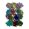
|
|---|---|
| 1 |
|
-Components
| #1: Protein | / 20S proteasome alpha subunit / Proteasome core protein PsmA Mass: 24776.281 Da / Num. of mol.: 14 Source method: isolated from a genetically manipulated source Source: (gene. exp.) Thermoplasma acidophilum (acidophilic) / Gene: psmA, Ta1288 / Production host:  Escherichia coli (E. coli) Escherichia coli (E. coli)References: UniProt: P25156, proteasome endopeptidase complex#2: Protein | / 20S proteasome beta subunit / Proteasome core protein PsmBMass: 22294.848 Da / Num. of mol.: 14 Source method: isolated from a genetically manipulated source Source: (gene. exp.) Thermoplasma acidophilum (acidophilic) / Gene: psmB, Ta0612 / Production host: Escherichia coli (E. coli)References: UniProt: P28061, proteasome endopeptidase complex |
|---|
-Experimental details
-Experiment
| Experiment | Method: ELECTRON MICROSCOPY |
|---|---|
| EM experiment | Aggregation state: PARTICLE / 3D reconstruction method: single particle reconstruction |
- Sample preparation
| Component |
| ||||||||||||||||
|---|---|---|---|---|---|---|---|---|---|---|---|---|---|---|---|---|---|
| Molecular weight | Value: 0.7 MDa / Experimental value: YES | ||||||||||||||||
| Specimen | Embedding applied: NO / Shadowing applied: NO / Staining applied: NO / Vitrification applied: YES | ||||||||||||||||
| Specimen support | Details: Quantifoil grid | ||||||||||||||||
| Vitrification | Instrument: FEI VITROBOT MARK III / Cryogen name: ETHANE / Temp: 120 K / Humidity: 100 % / Details: Plunged into liquid ethane (FEI VITROBOT MARK III) |
- Electron microscopy imaging
| Experimental equipment |  Model: Tecnai Polara / Image courtesy: FEI Company |
|---|---|
| Microscopy | Model: FEI POLARA 300 / Date: Jun 1, 2012 |
| Electron gun | Electron source: FIELD EMISSION GUN / Accelerating voltage: 300 kV / Illumination mode: FLOOD BEAM |
| Electron lens | Mode: BRIGHT FIELDBright-field microscopy / Nominal magnification: 31000 X / Nominal defocus max: 1900 nm / Nominal defocus min: 800 nm / Cs: 2 mm |
| Image recording | Electron dose: 20 e/Å2 / Film or detector model: GATAN K2 (4k x 4k) |
| Image scans | Num. digital images: 600 |
| Radiation wavelength | Relative weight: 1 |
- Processing
| EM software |
| ||||||||||||||||
|---|---|---|---|---|---|---|---|---|---|---|---|---|---|---|---|---|---|
| CTF correction | Details: Each Particle | ||||||||||||||||
| Symmetry | Point symmetry: D7 (2x7 fold dihedral) | ||||||||||||||||
| 3D reconstruction | Method: FREALIGN / Resolution: 3.3 Å / Resolution method: FSC 0.143 CUT-OFF / Num. of particles: 126729 / Nominal pixel size: 1.2156 Å / Actual pixel size: 1.2156 Å / Details: Gold Standard FSC, imposed symmetry: D7 / Refinement type: HALF-MAPS REFINED INDEPENDENTLY / Symmetry type: POINT | ||||||||||||||||
| Atomic model building | Protocol: FLEXIBLE FIT / Space: REAL / Details: REFINEMENT PROTOCOL--Flexible | ||||||||||||||||
| Atomic model building | PDB-ID: 1PMA Accession code: 1PMA / Source name: PDB / Type: experimental model | ||||||||||||||||
| Refinement step | Cycle: LAST
|
