Movie
Movie Controller

[English] 日本語
 Yorodumi
- PDB-3j6o: Kinetic and Structural Analysis of Coxsackievirus B3 Receptor Int... -
+ Open data

- Basic information
| Entry | Database: PDB / ID: 3j6o | ||||||
|---|---|---|---|---|---|---|---|
| Title | Kinetic and Structural Analysis of Coxsackievirus B3 Receptor Interactions and Formation of the A-particle | ||||||
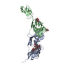
 Components Components | Coxsackie and adenovirus receptor | ||||||
 Keywords Keywords | CELL ADHESION / Coxsackievirus B3 / CVB3 / CAR | ||||||
| Function / homology |  Function and homology information Function and homology informationAV node cell-bundle of His cell adhesion involved in cell communication / cell adhesive protein binding involved in AV node cell-bundle of His cell communication / homotypic cell-cell adhesion / AV node cell to bundle of His cell communication / epithelial structure maintenance / regulation of AV node cell action potential / gamma-delta T cell activation / germ cell migration / apicolateral plasma membrane / transepithelial transport ...AV node cell-bundle of His cell adhesion involved in cell communication / cell adhesive protein binding involved in AV node cell-bundle of His cell communication / homotypic cell-cell adhesion / AV node cell to bundle of His cell communication / epithelial structure maintenance / regulation of AV node cell action potential / gamma-delta T cell activation / germ cell migration / apicolateral plasma membrane / transepithelial transport / cell-cell junction organization / connexin binding / cardiac muscle cell development / heterophilic cell-cell adhesion via plasma membrane cell adhesion molecules / intercalated disc / bicellular tight junction / cell adhesion molecule binding / neutrophil chemotaxis / acrosomal vesicle / mitochondrion organization / filopodium / PDZ domain binding / Cell surface interactions at the vascular wall / adherens junction / neuromuscular junction / beta-catenin binding / integrin binding / Immunoregulatory interactions between a Lymphoid and a non-Lymphoid cell / cell junction / cell-cell junction / virus receptor activity / heart development / basolateral plasma membrane / cell body / growth cone / actin cytoskeleton organization / defense response to virus / neuron projection / membrane raft / signaling receptor binding / protein-containing complex / extracellular space / extracellular region / nucleoplasm / identical protein binding / plasma membrane / cytoplasm Similarity search - Function | ||||||
| Biological species |  Homo sapiens (human) Homo sapiens (human) | ||||||
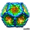
| Method | ELECTRON MICROSCOPY / single particle reconstruction / cryo EM / Resolution: 9 Å | ||||||
 Authors Authors | Organtini, L.J. / Makhov, A.M. / Conway, J.F. / Hafenstein, S. / Carson, S.D. | ||||||
 Citation Citation | Journal: J Virol / Year: 2014 Title: Kinetic and structural analysis of coxsackievirus B3 receptor interactions and formation of the A-particle. Authors: Lindsey J Organtini / Alexander M Makhov / James F Conway / Susan Hafenstein / Steven D Carson /  Abstract: The coxsackievirus and adenovirus receptor (CAR) has been identified as the cellular receptor for group B coxsackieviruses, including serotype 3 (CVB3). CAR mediates infection by binding to CVB3 and ...The coxsackievirus and adenovirus receptor (CAR) has been identified as the cellular receptor for group B coxsackieviruses, including serotype 3 (CVB3). CAR mediates infection by binding to CVB3 and catalyzing conformational changes in the virus that result in formation of the altered, noninfectious A-particle. Kinetic analyses show that the apparent first-order rate constant for the inactivation of CVB3 by soluble CAR (sCAR) at physiological temperatures varies nonlinearly with sCAR concentration. Cryo-electron microscopy (cryo-EM) reconstruction of the CVB3-CAR complex resulted in a 9.0-Å resolution map that was interpreted with the four available crystal structures of CAR, providing a consensus footprint for the receptor binding site. The analysis of the cryo-EM structure identifies important virus-receptor interactions that are conserved across picornavirus species. These conserved interactions map to variable antigenic sites or structurally conserved regions, suggesting a combination of evolutionary mechanisms for receptor site preservation. The CAR-catalyzed A-particle structure was solved to a 6.6-Å resolution and shows significant rearrangement of internal features and symmetric interactions with the RNA genome. IMPORTANCE: This report presents new information about receptor use by picornaviruses and highlights the importance of attaining at least an ∼9-Å resolution for the interpretation of cryo-EM ...IMPORTANCE: This report presents new information about receptor use by picornaviruses and highlights the importance of attaining at least an ∼9-Å resolution for the interpretation of cryo-EM complex maps. The analysis of receptor binding elucidates two complementary mechanisms for preservation of the low-affinity (initial) interaction of the receptor and defines the kinetics of receptor-catalyzed conformational change to the A-particle. | ||||||
| History |
|
- Structure visualization
| Movie |
Movie viewer |
|---|---|
| Structure viewer | Molecule: MolmilJmol/JSmol |
- Downloads & links
-Download
| PDBx/mmCIF format | 3j6o.cif.gz | 46.9 KB | Display | PDBx/mmCIF format |
|---|---|---|---|---|
| PDB format | pdb3j6o.ent.gz | 34.9 KB | Display | PDB format |
| PDBx/mmJSON format | 3j6o.json.gz | Tree view | PDBx/mmJSON format | |
| Others |  Other downloads Other downloads |
-Validation report
| Summary document | 3j6o_validation.pdf.gz | 757.4 KB | Display | wwPDB validaton report |
|---|---|---|---|---|
| Full document | 3j6o_full_validation.pdf.gz | 762.8 KB | Display | |
| Data in XML | 3j6o_validation.xml.gz | 14.6 KB | Display | |
| Data in CIF | 3j6o_validation.cif.gz | 19.3 KB | Display | |
| Arichive directory | https://data.pdbj.org/pub/pdb/validation_reports/j6/3j6oftp://data.pdbj.org/pub/pdb/validation_reports/j6/3j6o | HTTPS FTP |
-Related structure data
| Related structure data | 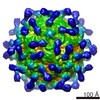 5927MC 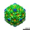 5928C 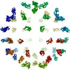 3j6lC 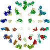 3j6mC 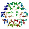 3j6nC M: map data used to model this data C: citing same article ( |
|---|---|
| Similar structure data |
-Links
 PDBj
PDBj


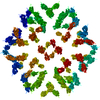
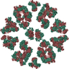
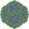
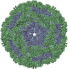
- Assembly
| Deposited unit | 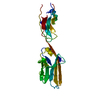
|
|---|---|
| 1 | x 60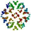
|
| 2 |
|
| 3 | x 5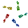
|
| 4 | x 6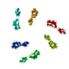
|
| 5 | 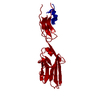
|
| Symmetry | Point symmetry: (Schoenflies symbol: I (icosahedral)) |
-Components
| #1: Protein | Mass: 25046.229 Da / Num. of mol.: 1 / Fragment: SEE REMARK 999 Source method: isolated from a genetically manipulated source Source: (gene. exp.) Homo sapiens (human) / Production host:  |
|---|---|
| Sequence details | THE HUMAN PROTEIN WAS IMAGED, BUT THE PROTEIN SEQUENCE THAT WAS FIT TO THE ELECTRON MICROSCOPY MAP ...THE HUMAN PROTEIN WAS IMAGED, BUT THE PROTEIN SEQUENCE THAT WAS FIT TO THE ELECTRON MICROSCOPY |
-Experimental details
-Experiment
| Experiment | Method: ELECTRON MICROSCOPY |
|---|---|
| EM experiment | Aggregation state: PARTICLE / 3D reconstruction method: single particle reconstruction |
- Sample preparation
| Component |
| ||||||||||||||||||||
|---|---|---|---|---|---|---|---|---|---|---|---|---|---|---|---|---|---|---|---|---|---|
| Molecular weight | Value: 7 MDa / Experimental value: YES | ||||||||||||||||||||
| Details of virus | Empty: NO / Enveloped: NO / Host category: VERTEBRATES / Isolate: STRAIN / Type: VIRION | ||||||||||||||||||||
| Natural host | Organism: Homo sapiens | ||||||||||||||||||||
| Buffer solution | Name: 50 mM MES, 100 mM NaCl / pH: 6 / Details: 50 mM MES, 100 mM NaCl | ||||||||||||||||||||
| Specimen | Conc.: 0.1 mg/ml / Embedding applied: NO / Shadowing applied: NO / Staining applied: NO / Vitrification applied: YES | ||||||||||||||||||||
| Specimen support | Details: glow-discharged holey carbon Quantifoil electron microscopy grids | ||||||||||||||||||||
| Vitrification | Instrument: FEI VITROBOT MARK III / Cryogen name: OTHER / Temp: 95 K / Humidity: 95 % Details: Plunged into ethane-propane (FEI VITROBOT MARK III) |
- Electron microscopy imaging
| Experimental equipment |  Model: Tecnai F20 / Image courtesy: FEI Company |
|---|---|
| Microscopy | Model: FEI TECNAI F20 / Date: Aug 1, 2012 |
| Electron gun | Electron source:  FIELD EMISSION GUN / Accelerating voltage: 200 kV / Illumination mode: SPOT SCAN FIELD EMISSION GUN / Accelerating voltage: 200 kV / Illumination mode: SPOT SCAN |
| Electron lens | Mode: BRIGHT FIELD / Nominal magnification: 50000 X / Calibrated magnification: 50000 X / Nominal defocus max: 3660 nm / Nominal defocus min: 1980 nm / Cs: 2 mm / Astigmatism: CTFFIND3 / Camera length: 0 mm |
| Specimen holder | Specimen holder model: GATAN LIQUID NITROGEN / Tilt angle max: 0 ° / Tilt angle min: 0 ° |
| Image recording | Electron dose: 15 e/Å2 / Film or detector model: KODAK SO-163 FILM |
| Image scans | Num. digital images: 96 |
| Radiation | Protocol: SINGLE WAVELENGTH / Monochromatic (M) / Laue (L): M / Scattering type: x-ray |
| Radiation wavelength | Relative weight: 1 |
- Processing
| EM software |
| ||||||||||||
|---|---|---|---|---|---|---|---|---|---|---|---|---|---|
| CTF correction | Details: AUTO3DEM | ||||||||||||
| Symmetry | Point symmetry: I (icosahedral) | ||||||||||||
| 3D reconstruction | Method: Common Lines / Resolution: 9 Å / Resolution method: FSC 0.5 CUT-OFF / Num. of particles: 9302 / Nominal pixel size: 1.25 Å / Actual pixel size: 1.25 Å Details: (Single particle details: Particles were selected using EMAN) (Single particle--Applied symmetry: I) Symmetry type: POINT | ||||||||||||
| Atomic model building | Protocol: RIGID BODY FIT / Space: REAL / Target criteria: correlation coefficient / Details: REFINEMENT PROTOCOL--rigid body | ||||||||||||
| Atomic model building | PDB-ID: 3MJ7 Pdb chain-ID: S | ||||||||||||
| Refinement step | Cycle: LAST
|