Movie
Movie Controller


+ Open data

- Basic information
| Entry | Database: PDB / ID: 3j24 | ||||||
|---|---|---|---|---|---|---|---|
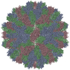
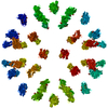
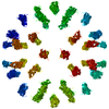
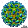
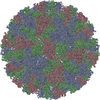
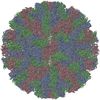
| Title | CryoEM reconstruction of complement decay-accelerating factor | ||||||
 Components Components | Complement decay-accelerating factor | ||||||
 Keywords Keywords |  IMMUNE SYSTEM / blood group antigen / complement pathway / glycoprotein / GPI-anchor / immune response / innate immunity / lipoprotein / membrane / Sushi IMMUNE SYSTEM / blood group antigen / complement pathway / glycoprotein / GPI-anchor / immune response / innate immunity / lipoprotein / membrane / Sushi | ||||||
| Function / homology |  Function and homology information Function and homology informationnegative regulation of complement activation / regulation of lipopolysaccharide-mediated signaling pathway / regulation of complement-dependent cytotoxicity / regulation of complement activation / respiratory burst / positive regulation of CD4-positive, alpha-beta T cell activation / positive regulation of CD4-positive, alpha-beta T cell proliferation / Class B/2 (Secretin family receptors) / ficolin-1-rich granule membrane / side of membrane ...negative regulation of complement activation / regulation of lipopolysaccharide-mediated signaling pathway / regulation of complement-dependent cytotoxicity / regulation of complement activation / respiratory burst / positive regulation of CD4-positive, alpha-beta T cell activation / positive regulation of CD4-positive, alpha-beta T cell proliferation / Class B/2 (Secretin family receptors) / ficolin-1-rich granule membrane / side of membrane / COPI-mediated anterograde transport / complement activation, classical pathway / transport vesicle / endoplasmic reticulum-Golgi intermediate compartment membrane / secretory granule membrane / Regulation of Complement cascade / positive regulation of T cell cytokine production / virus receptor activity / positive regulation of cytosolic calcium ion concentration / membrane raft / Golgi membrane / innate immune response / lipid binding / Neutrophil degranulation / cell surface / extracellular exosome / extracellular region / plasma membraneSimilarity search - Function | ||||||
| Biological species |  Homo sapiens (human) Homo sapiens (human) | ||||||
| Method | ELECTRON MICROSCOPY / single particle reconstruction / cryo EM / Resolution: 9 Å | ||||||
 Authors Authors | Yoder, J.D. / Hafenstein, S.H. | ||||||
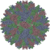
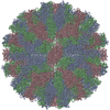
 Citation Citation | Journal: J Virol / Year: 2012 Title: The crystal structure of a coxsackievirus B3-RD variant and a refined 9-angstrom cryo-electron microscopy reconstruction of the virus complexed with decay-accelerating factor (DAF) provide a ...Title: The crystal structure of a coxsackievirus B3-RD variant and a refined 9-angstrom cryo-electron microscopy reconstruction of the virus complexed with decay-accelerating factor (DAF) provide a new footprint of DAF on the virus surface. Authors: Joshua D Yoder / Javier O Cifuente / Jieyan Pan / Jeffrey M Bergelson / Susan Hafenstein /  Abstract: The coxsackievirus-adenovirus receptor (CAR) and decay-accelerating factor (DAF) have been identified as cellular receptors for coxsackievirus B3 (CVB3). The first described DAF-binding isolate was ...The coxsackievirus-adenovirus receptor (CAR) and decay-accelerating factor (DAF) have been identified as cellular receptors for coxsackievirus B3 (CVB3). The first described DAF-binding isolate was obtained during passage of the prototype strain, Nancy, on rhabdomyosarcoma (RD) cells, which express DAF but very little CAR. Here, the structure of the resulting variant, CVB3-RD, has been solved by X-ray crystallography to 2.74 Å, and a cryo-electron microscopy reconstruction of CVB3-RD complexed with DAF has been refined to 9.0 Å. This new high-resolution structure permits us to correct an error in our previous view of DAF-virus interactions, providing a new footprint of DAF that bridges two adjacent protomers. The contact sites between the virus and DAF clearly encompass CVB3-RD residues recently shown to be required for binding to DAF; these residues interact with DAF short consensus repeat 2 (SCR2), which is known to be essential for virus binding. Based on the new structure, the mode of the DAF interaction with CVB3 differs significantly from the mode reported previously for DAF binding to echoviruses. | ||||||
| History |
|
- Structure visualization
| Movie |
Movie viewer |
|---|---|
| Structure viewer | Molecule: MolmilJmol/JSmol |
- Downloads & links
-Download
| PDBx/mmCIF format | 3j24.cif.gz | 57.3 KB | Display | PDBx/mmCIF format |
|---|---|---|---|---|
| PDB format | pdb3j24.ent.gz | 44.6 KB | Display | PDB format |
| PDBx/mmJSON format | 3j24.json.gz | Tree view | PDBx/mmJSON format | |
| Others |  Other downloads Other downloads |
-Validation report
| Arichive directory | https://data.pdbj.org/pub/pdb/validation_reports/j2/3j24ftp://data.pdbj.org/pub/pdb/validation_reports/j2/3j24 | HTTPS FTP |
|---|
-Related structure data
| Related structure data | 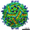 5475MC  4gb3C M: map data used to model this data C: citing same article ( |
|---|---|
| Similar structure data |
-Links
 PDBj
PDBj


- Assembly
| Deposited unit | 
|
|---|---|
| 1 | x 60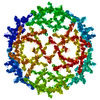
|
| 2 |
|
| 3 | x 5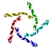
|
| 4 | x 6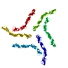
|
| 5 | 
|
| Symmetry | Point symmetry: (Schoenflies symbol: I (icosahedral)) |
-Components
| #1: Protein | Mass: 28174.666 Da / Num. of mol.: 1 Fragment: four extracellular SCR domains (UNP residues 35-285) Source method: isolated from a genetically manipulated source Source: (gene. exp.) Homo sapiens (human) / Gene: CD55, CR, DAF / Production host:  Escherichia coli (E. coli) / References: UniProt: P08174 Escherichia coli (E. coli) / References: UniProt: P08174 |
|---|
-Experimental details
-Experiment
| Experiment | Method: ELECTRON MICROSCOPY |
|---|---|
| EM experiment | Aggregation state: PARTICLE / 3D reconstruction method: single particle reconstruction |
- Sample preparation
| Component | Name: Complement decay-accelerating factor bound to coxsackievirus B3-RD strain Type: VIRUS |
|---|---|
| Buffer solution | Name: 2-(N-morpholino)ethanesulfonic acid (MES) / pH: 6 / Details: 50mM MES |
| Specimen | Conc.: 2 mg/ml / Embedding applied: NO / Shadowing applied: NO / Staining applied: NO / Vitrification applied: YES |
| Specimen support | Details: Quantifoil |
| Vitrification | Instrument: HOMEMADE PLUNGER / Cryogen name: ETHANE / Temp: 120 K Details: Blotted before plunging in liquid ethane (homemade plunger). |
- Electron microscopy imaging
| Microscopy | Model: FEI/PHILIPS CM300FEG/T / Date: Aug 4, 2006 |
|---|---|
| Electron gun | Electron source: TUNGSTEN HAIRPIN / Accelerating voltage: 300 kV / Illumination mode: FLOOD BEAM |
| Electron lens | Mode: BRIGHT FIELDBright-field microscopy / Nominal magnification: 45000 X / Calibrated magnification: 47000 X / Nominal defocus max: 4600 nm / Nominal defocus min: 1000 nm / Cs: 2 mm |
| Specimen holder | Specimen holder model: GATAN LIQUID NITROGEN / Specimen holder type: Side mounted nitrogen cooled / Temperature: 93 K / Temperature (max): 93 K / Temperature (min): 83 K / Tilt angle max: 0 ° / Tilt angle min: 0 ° |
| Image recording | Electron dose: 24 e/Å2 / Film or detector model: KODAK SO-163 FILM |
| Image scans | Num. digital images: 36 |
| Radiation | Protocol: SINGLE WAVELENGTH / Monochromatic (M) / Laue (L): M / Scattering type: x-ray |
| Radiation wavelength | Relative weight: 1 |
- Processing
| EM software |
| ||||||||||||||||||
|---|---|---|---|---|---|---|---|---|---|---|---|---|---|---|---|---|---|---|---|
| CTF correction | Details: AUTO3DEM | ||||||||||||||||||
| Symmetry | Point symmetry: I (icosahedral) | ||||||||||||||||||
| 3D reconstruction | Method: common lines / Resolution: 9 Å / Num. of particles: 3010 / Nominal pixel size: 2.94 Å / Actual pixel size: 2.94 Å / Symmetry type: POINT | ||||||||||||||||||
| Atomic model building | Protocol: RIGID BODY FIT / Space: REAL / Target criteria: average map value Details: METHOD--Local refinement, rigid body REFINEMENT PROTOCOL--rigid body | ||||||||||||||||||
| Atomic model building | PDB-ID: 1OJW Pdb chain-ID: B | ||||||||||||||||||
| Refinement step | Cycle: LAST
|