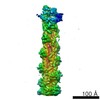
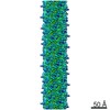
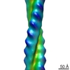
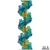
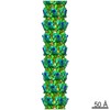
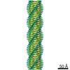
Journal: Proc Natl Acad Sci U S A / Year: 2011 Title: Remodeling of actin filaments by ADF/cofilin proteins. Authors: Vitold E Galkin / Albina Orlova / Dmitri S Kudryashov / Alexander Solodukhin / Emil Reisler / Gunnar F Schröder / Edward H Egelman / Abstract: Cofilin/ADF proteins play key roles in the dynamics of actin, one of the most abundant and highly conserved eukaryotic proteins. We used cryoelectron microscopy to generate a 9-Å resolution three- ...Cofilin/ADF proteins play key roles in the dynamics of actin, one of the most abundant and highly conserved eukaryotic proteins. We used cryoelectron microscopy to generate a 9-Å resolution three-dimensional reconstruction of cofilin-decorated actin filaments, the highest resolution achieved for a complex of F-actin with an actin-binding protein. We show that the cofilin-induced change in the filament twist is due to a unique conformation of the actin molecule unrelated to any previously observed state. The changes between the actin protomer in naked F-actin and in the actin-cofilin filament are greater than the conformational changes between G- and F-actin. Our results show the structural plasticity of actin, suggest that other actin-binding proteins may also induce large but different conformational changes, and show that F-actin cannot be described by a single molecular model.
THE ASSEMBLY REPRESENTED IN THIS ENTRY HAS REGULAR HELICAL SYMMETRY WITH THE FOLLOWING PARAMETERS: ROTATION PER SUBUNIT (TWIST) = -162.1 DEGREES; RISE PER SUBUNIT (HEIGHT) = 27.6 ANGSTROM
-
Components
#1: Protein
Actin, cytoplasmic1 / Beta-actin
Mass: 41651.465 Da / Num. of mol.: 12 / Source method: isolated from a natural source / Source: (natural) Gallus gallus (chicken) / Tissue: muscle / References: UniProt: P60706
#2: Protein
Cofilin-2 / Cofilin / muscle isoform
Mass: 18532.531 Da / Num. of mol.: 12 / Fragment: SEE REMARK 999 Source method: isolated from a genetically manipulated source Source: (gene. exp.) Homo sapiens (human) / Gene: CFL1, CFL / Production host: Escherichia coli (E. coli) / References: UniProt: P23528*PLUS
Sequence details
THE AUTHORS STATE THAT HUMAN COFILIN-2 WAS USED IN THE EXPERIMENT, BUT HUMAN COFILIN-1 (UNP P23528) ...THE AUTHORS STATE THAT HUMAN COFILIN-2 WAS USED IN THE EXPERIMENT, BUT HUMAN COFILIN-1 (UNP P23528) WAS USED FOR MODELING.
-
Experimental details
-
Experiment
Experiment
Method: ELECTRON MICROSCOPY
EM experiment
Aggregation state: FILAMENT / 3D reconstruction method: helical reconstruction
-
Sample preparation
Component
Name: actin decorated with cofilin / Type: COMPLEX / Details: filament containing one cofilin to one actin
Specimen
Embedding applied: NO / Shadowing applied: NO / Staining applied: NO / Vitrification applied: YES
Vitrification
Instrument: FEI VITROBOT MARK I / Cryogen name: ETHANE / Humidity: 100 %
-
Electron microscopy imaging
Experimental equipment
Model: Tecnai F20 / Image courtesy: FEI Company
Microscopy
Model: FEI TECNAI F20 / Date: Jan 1, 2010
Electron gun
Electron source: FIELD EMISSION GUN / Accelerating voltage: 200 kV / Illumination mode: FLOOD BEAM
Electron lens
Mode: BRIGHT FIELD / Nominal magnification: 50000 X / Nominal defocus max: 5300 nm / Nominal defocus min: 1100 nm / Cs: 2 mm
In the structure databanks used in Yorodumi, some data are registered as the other names, "COVID-19 virus" and "2019-nCoV". Here are the details of the virus and the list of structure data.
Jan 31, 2019. EMDB accession codes are about to change! (news from PDBe EMDB page)
EMDB accession codes are about to change! (news from PDBe EMDB page)
The allocation of 4 digits for EMDB accession codes will soon come to an end. Whilst these codes will remain in use, new EMDB accession codes will include an additional digit and will expand incrementally as the available range of codes is exhausted. The current 4-digit format prefixed with “EMD-” (i.e. EMD-XXXX) will advance to a 5-digit format (i.e. EMD-XXXXX), and so on. It is currently estimated that the 4-digit codes will be depleted around Spring 2019, at which point the 5-digit format will come into force.
The EM Navigator/Yorodumi systems omit the EMD- prefix.
Related info.:Q: What is EMD? / ID/Accession-code notation in Yorodumi/EM Navigator
Yorodumi is a browser for structure data from EMDB, PDB, SASBDB, etc.
This page is also the successor to EM Navigator detail page, and also detail information page/front-end page for Omokage search.
The word "yorodu" (or yorozu) is an old Japanese word meaning "ten thousand". "mi" (miru) is to see.
Related info.:EMDB / PDB / SASBDB / Comparison of 3 databanks / Yorodumi Search / Aug 31, 2016. New EM Navigator & Yorodumi / Yorodumi Papers / Jmol/JSmol / Function and homology information / Changes in new EM Navigator and Yorodumi
 Movie
Movie Controller


 Open data
 Basic information
 Components
 Keywords
 Function and homology information
 Homo sapiens (human)
 Authors
 Citation
 Structure visualization
 Downloads & links
 Other downloads
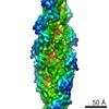
 PDBj
PDBj


 Assembly
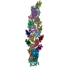

 Sample preparation
 Electron microscopy imaging
 FIELD EMISSION GUN / Accelerating voltage: 200 kV / Illumination mode: FLOOD BEAM
 Processing