 Movie
Movie Controller


+ Open data

- Basic information
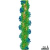
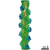
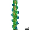
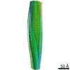
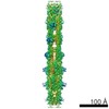
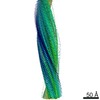
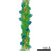
| Entry | Database: PDB / ID: 3g37 | ||||||
|---|---|---|---|---|---|---|---|
| Title | Cryo-EM structure of actin filament in the presence of phosphate | ||||||
 Components Components | Actin, alpha skeletal muscle | ||||||
 Keywords Keywords | CONTRACTILE PROTEIN / actin / cytoskeleton / cell adhesion / cellular signaling / cytokinesis / muscle / cryo-EM / ATP-binding / Methylation / Muscle protein / Nucleotide-binding / Phosphoprotein | ||||||
| Function / homology |  Function and homology information Function and homology informationcytoskeletal motor activator activity / tropomyosin binding / mesenchyme migration / troponin I binding / myosin heavy chain binding / filamentous actin / actin filament bundle / skeletal muscle thin filament assembly / striated muscle thin filament / actin filament bundle assembly ...cytoskeletal motor activator activity / tropomyosin binding / mesenchyme migration / troponin I binding / myosin heavy chain binding / filamentous actin / actin filament bundle / skeletal muscle thin filament assembly / striated muscle thin filament / actin filament bundle assembly / skeletal muscle myofibril / actin monomer binding / skeletal muscle fiber development / stress fiber / titin binding / actin filament polymerization / filopodium / actin filament / Hydrolases; Acting on acid anhydrides; Acting on acid anhydrides to facilitate cellular and subcellular movement / calcium-dependent protein binding / lamellipodium / cell body / hydrolase activity / protein domain specific binding / calcium ion binding / positive regulation of gene expression / magnesium ion binding / ATP binding / identical protein binding / cytoplasm Similarity search - Function | ||||||
| Biological species |  | ||||||
| Method | ELECTRON MICROSCOPY / single particle reconstruction / cryo EM / Resolution: 6 Å | ||||||
 Authors Authors | Wakabayshi, T. / Murakami, K. / Yasunaga, T. / Noguchi, T.Q. / Uyeda, T.Q. | ||||||
 Citation Citation | Journal: Cell / Year: 2010 Title: Structural basis for actin assembly, activation of ATP hydrolysis, and delayed phosphate release. Authors: Kenji Murakami / Takuo Yasunaga / Taro Q P Noguchi / Yuki Gomibuchi / Kien X Ngo / Taro Q P Uyeda / Takeyuki Wakabayashi /  Abstract: Assembled actin filaments support cellular signaling, intracellular trafficking, and cytokinesis. ATP hydrolysis triggered by actin assembly provides the structural cues for filament turnover in vivo. ...Assembled actin filaments support cellular signaling, intracellular trafficking, and cytokinesis. ATP hydrolysis triggered by actin assembly provides the structural cues for filament turnover in vivo. Here, we present the cryo-electron microscopic (cryo-EM) structure of filamentous actin (F-actin) in the presence of phosphate, with the visualization of some α-helical backbones and large side chains. A complete atomic model based on the EM map identified intermolecular interactions mediated by bound magnesium and phosphate ions. Comparison of the F-actin model with G-actin monomer crystal structures reveals a critical role for bending of the conserved proline-rich loop in triggering phosphate release following ATP hydrolysis. Crystal structures of G-actin show that mutations in this loop trap the catalytic site in two intermediate states of the ATPase cycle. The combined structural information allows us to propose a detailed molecular mechanism for the biochemical events, including actin polymerization and ATPase activation, critical for actin filament dynamics. | ||||||
| History |
|
- Structure visualization
| Movie |
Movie viewer |
|---|---|
| Structure viewer | Molecule: MolmilJmol/JSmol |
- Downloads & links
-Download
| PDBx/mmCIF format | 3g37.cif.gz | 759 KB | Display | PDBx/mmCIF format |
|---|---|---|---|---|
| PDB format | pdb3g37.ent.gz | 631.8 KB | Display | PDB format |
| PDBx/mmJSON format | 3g37.json.gz | Tree view | PDBx/mmJSON format | |
| Others |  Other downloads Other downloads |
-Validation report
| Summary document | 3g37_validation.pdf.gz | 1.6 MB | Display | wwPDB validaton report |
|---|---|---|---|---|
| Full document | 3g37_full_validation.pdf.gz | 1.7 MB | Display | |
| Data in XML | 3g37_validation.xml.gz | 131.7 KB | Display | |
| Data in CIF | 3g37_validation.cif.gz | 191.1 KB | Display | |
| Arichive directory | https://data.pdbj.org/pub/pdb/validation_reports/g3/3g37ftp://data.pdbj.org/pub/pdb/validation_reports/g3/3g37 | HTTPS FTP |
-Related structure data
| Related structure data |  1674MC 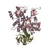 3a5lC 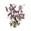 3a5mC 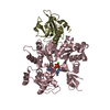 3a5nC 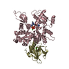 3a5oC M: map data used to model this data C: citing same article ( |
|---|---|
| Similar structure data |

-Links
 PDBj
PDBj


- Assembly
| Deposited unit | 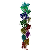
|
|---|---|
| 1 |
|
-Components
| #1: Protein | Mass: 41901.668 Da / Num. of mol.: 12 / Source method: isolated from a natural source / Source: (natural) #2: Chemical | ChemComp-ADP /   Mass: 427.201 Da / Num. of mol.: 12 / Source method: obtained synthetically / Formula: C10H15N5O10P2 / Comment: ADP, energy-carrying molecule*YM Mass: 427.201 Da / Num. of mol.: 12 / Source method: obtained synthetically / Formula: C10H15N5O10P2 / Comment: ADP, energy-carrying molecule*YM#3: Chemical | ChemComp-PO4 /   Mass: 94.971 Da / Num. of mol.: 36 / Source method: obtained synthetically / Formula: PO4 Mass: 94.971 Da / Num. of mol.: 36 / Source method: obtained synthetically / Formula: PO4#4: Chemical | ChemComp-MG /   Mass: 24.305 Da / Num. of mol.: 72 / Source method: obtained synthetically / Formula: Mg Mass: 24.305 Da / Num. of mol.: 72 / Source method: obtained synthetically / Formula: Mg |
|---|
-Experimental details
-Experiment
| Experiment | Method: ELECTRON MICROSCOPY |
|---|---|
| EM experiment | Aggregation state: FILAMENT / 3D reconstruction method: single particle reconstruction |
- Sample preparation
| Component | Name: actin filament in the presence of phosphate / Type: COMPLEX Details: 50 mM NaCl, 5 mM MgCl2, 0.025 mM ATP, 10 mM sodium phosphate (pH 7.4), 0.05 %NaN3, 0.7 mM DTT |
|---|---|
| Buffer solution | Name: phosphate buffer / pH: 7.4 / Details: phosphate buffer |
| Specimen | Embedding applied: NO / Shadowing applied: NO / Staining applied: NO / Vitrification applied: YES |
| Vitrification | Cryogen name: ETHANE / Temp: 77 K / Humidity: 90 % |
- Electron microscopy imaging
| Microscopy | Model: HITACHI EF2000 |
|---|---|
| Electron gun | Electron source:  FIELD EMISSION GUN / Accelerating voltage: 200 kV / Illumination mode: SPOT SCAN FIELD EMISSION GUN / Accelerating voltage: 200 kV / Illumination mode: SPOT SCAN |
| Electron lens | Mode: BRIGHT FIELD / Nominal magnification: 100000 X / Nominal defocus max: 1500 nm / Nominal defocus min: 1000 nm |
| Specimen holder | Specimen holder model: GATAN LIQUID NITROGEN Specimen holder type: Side entry liquid nitrogen-cooled cryo specimen holder Temperature: 77 K / Temperature (max): 77 K / Temperature (min): 77 K / Tilt angle max: 0 ° / Tilt angle min: 0 ° |
| Image recording | Electron dose: 15 e/Å2 / Film or detector model: GENERIC CCD Details: CCD camera with an epitaxially-grown CsI scintillator |
| EM imaging optics | Energyfilter name: In-column Omega Filter |
- Processing
| EM software |
| ||||||||||||||||
|---|---|---|---|---|---|---|---|---|---|---|---|---|---|---|---|---|---|
| CTF correction | Details: FSC at 0.143 cut-off | ||||||||||||||||
| 3D reconstruction | Method: single particle / Resolution: 6 Å / Num. of particles: 8000 / Nominal pixel size: 2.275 Å / Actual pixel size: 2.275 Å / Symmetry type: HELICAL | ||||||||||||||||
| Atomic model building | Protocol: FLEXIBLE FIT / Space: REAL / Target criteria: pdbRhofit (Yasunaga & Wakabayashi, 1996) Details: METHOD--local refinement REFINEMENT PROTOCOL--real-space refinement | ||||||||||||||||
| Refinement step | Cycle: LAST
|
