MEMBRANE PROTEIN / membrane protein sodium proton antiporter / Antiport / Cell inner membrane / Cell membrane / Ion transport / Membrane / Sodium transport / Transmembrane / Transport
Function / homology
Function and homology information
response to alkaline pH / sodium:proton antiporter activity / cardiolipin binding / response to salt stress / regulation of intracellular pH / plasma membrane Similarity search - Function
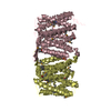
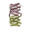
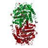
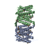
Journal: J Mol Biol / Year: 2009 Title: Conformations of NhaA, the Na/H exchanger from Escherichia coli, in the pH-activated and ion-translocating states. Authors: Matthias Appel / Dilem Hizlan / Kutti R Vinothkumar / Christine Ziegler / Werner Kühlbrandt / Abstract: NhaA, the main sodium-proton exchanger in the inner membrane of Escherichia coli, regulates the cytosolic concentrations of H and Na. It is inactive at acidic pH, becomes active between pH 6 and pH ...NhaA, the main sodium-proton exchanger in the inner membrane of Escherichia coli, regulates the cytosolic concentrations of H and Na. It is inactive at acidic pH, becomes active between pH 6 and pH 7, and reaches maximum activity at pH 8. By cryo-electron microscopy of two-dimensional crystals grown at pH 4 and incubated at higher pH, we identified two sequential conformational changes in the protein in response to pH or substrate ions. The first change is induced by a rise in pH from 6 to 7 and marks the transition from the inactive state to the pH-activated state. pH activation, which precedes the ion-induced conformational change, is accompanied by an overall expansion of the NhaA monomer and a local ordering of the N-terminus. The second conformational change is induced by the substrate ions Na and Li at pH above 7 and involves a 7-A displacement of helix IVp. This movement would cause a charge imbalance at the ion-binding site that may trigger the release of the substrate ion and open a periplasmic exit channel.
In the structure databanks used in Yorodumi, some data are registered as the other names, "COVID-19 virus" and "2019-nCoV". Here are the details of the virus and the list of structure data.
Jan 31, 2019. EMDB accession codes are about to change! (news from PDBe EMDB page)
EMDB accession codes are about to change! (news from PDBe EMDB page)
The allocation of 4 digits for EMDB accession codes will soon come to an end. Whilst these codes will remain in use, new EMDB accession codes will include an additional digit and will expand incrementally as the available range of codes is exhausted. The current 4-digit format prefixed with “EMD-” (i.e. EMD-XXXX) will advance to a 5-digit format (i.e. EMD-XXXXX), and so on. It is currently estimated that the 4-digit codes will be depleted around Spring 2019, at which point the 5-digit format will come into force.
The EM Navigator/Yorodumi systems omit the EMD- prefix.
Related info.:Q: What is EMD? / ID/Accession-code notation in Yorodumi/EM Navigator
Yorodumi is a browser for structure data from EMDB, PDB, SASBDB, etc.
This page is also the successor to EM Navigator detail page, and also detail information page/front-end page for Omokage search.
The word "yorodu" (or yorozu) is an old Japanese word meaning "ten thousand". "mi" (miru) is to see.
Related info.:EMDB / PDB / SASBDB / Comparison of 3 databanks / Yorodumi Search / Aug 31, 2016. New EM Navigator & Yorodumi / Yorodumi Papers / Jmol/JSmol / Function and homology information / Changes in new EM Navigator and Yorodumi
 Movie
Movie Controller


 Open data
 Basic information
 Components
 Keywords
 Function and homology information
 Authors
 Citation
 Structure visualization
 Downloads & links
 Other downloads


 PDBj
PDBj
 Assembly

 Sample preparation
 FIELD EMISSION GUN / Accelerating voltage: 300 kV / Illumination mode: FLOOD BEAM
 Processing