[English] 日本語
Yorodumi
- PDB-3dco: Drosophila NOD (3DC4) and Bovine Tubulin (1JFF) Docked into the 1... -

+
Open data


ID or keywords:

Loading...

-
Basic information

Entry
Database: PDB / ID: 3dco
TitleDrosophila NOD (3DC4) and Bovine Tubulin (1JFF) Docked into the 11-Angstrom Cryo-EM Map of Nucleotide-Free NOD Complexed to the Microtubule
Components
  • Bovine Alpha Tubulin
  • Bovine Beta Tubulin
  • Kinesin-like protein Nod
KeywordsMOTOR PROTEIN / kinesin / catalytic domain / ATPase / microtubule / ADP / nucleotide-binding protein / cryo-EM / 3D reconstruction / tubulin / nucleotide-free / ATP-binding / Coiled coil / Nucleotide-binding
Function / homology
Function and homology information


establishment of meiotic spindle orientation / distributive segregation / COPI-dependent Golgi-to-ER retrograde traffic / Kinesins / meiotic chromosome segregation / female meiotic nuclear division / microtubule plus-end binding / spindle assembly involved in female meiosis I / positive regulation of axon guidance / kinesin complex ...establishment of meiotic spindle orientation / distributive segregation / COPI-dependent Golgi-to-ER retrograde traffic / Kinesins / meiotic chromosome segregation / female meiotic nuclear division / microtubule plus-end binding / spindle assembly involved in female meiosis I / positive regulation of axon guidance / kinesin complex / microtubule motor activity / microtubule-based movement / cytoplasmic microtubule / microtubule-based process / positive regulation of microtubule polymerization / cellular response to interleukin-4 / Hydrolases; Acting on acid anhydrides; Acting on GTP to facilitate cellular and subcellular movement / structural constituent of cytoskeleton / microtubule cytoskeleton organization / microtubule cytoskeleton / double-stranded RNA binding / mitotic cell cycle / nervous system development / microtubule binding / microtubule / protein heterodimerization activity / GTPase activity / ubiquitin protein ligase binding / GTP binding / ATP hydrolysis activity / ATP binding / metal ion binding / cytosol / cytoplasm
Similarity search - Function
Kinesin-like protein / RuvA domain 2-like / Kinesin motor domain signature. / Kinesin motor domain, conserved site / Kinesin motor domain / Kinesin motor domain profile. / Kinesin motor, catalytic domain. ATPase. / Kinesin motor domain / Kinesin motor domain superfamily / Tubulin-beta mRNA autoregulation signal. ...Kinesin-like protein / RuvA domain 2-like / Kinesin motor domain signature. / Kinesin motor domain, conserved site / Kinesin motor domain / Kinesin motor domain profile. / Kinesin motor, catalytic domain. ATPase. / Kinesin motor domain / Kinesin motor domain superfamily / Tubulin-beta mRNA autoregulation signal. / Alpha tubulin / Beta tubulin, autoregulation binding site / Beta tubulin / Tubulin / Tubulin, C-terminal / Tubulin C-terminal domain / Tubulin, conserved site / Tubulin subunits alpha, beta, and gamma signature. / Tubulin/FtsZ family, C-terminal domain / Tubulin/FtsZ-like, C-terminal domain / Tubulin/FtsZ, C-terminal / Tubulin/FtsZ, 2-layer sandwich domain / Tubulin/FtsZ family, GTPase domain / Tubulin/FtsZ family, GTPase domain / Tubulin/FtsZ, GTPase domain / Tubulin/FtsZ, GTPase domain superfamily / P-loop containing nucleoside triphosphate hydrolase
Similarity search - Domain/homology
ADENOSINE-5'-DIPHOSPHATE / GUANOSINE-5'-DIPHOSPHATE / GUANOSINE-5'-TRIPHOSPHATE / TAXOL / Kinesin-like protein Nod / Tubulin alpha-1B chain / Tubulin beta-2B chain
Similarity search - Component
Biological speciesDrosophila melanogaster (fruit fly)
Bos taurus (cattle)
MethodELECTRON MICROSCOPY / SYNCHROTRON / single particle reconstruction / cryo EM / Resolution: 11 Å
AuthorsSindelar, C.V. / Cochran, J.C. / Kull, F.J.
Citation
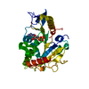
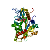
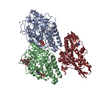
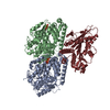
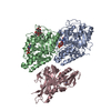
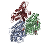
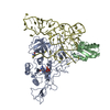
Journal: Cell / Year: 2009
Title: ATPase cycle of the nonmotile kinesin NOD allows microtubule end tracking and drives chromosome movement.
Authors: Jared C Cochran / Charles V Sindelar / Natasha K Mulko / Kimberly A Collins / Stephanie E Kong / R Scott Hawley / F Jon Kull /
Abstract: Segregation of nonexchange chromosomes during Drosophila melanogaster meiosis requires the proper function of NOD, a nonmotile kinesin-10. We have determined the X-ray crystal structure of the NOD ...Segregation of nonexchange chromosomes during Drosophila melanogaster meiosis requires the proper function of NOD, a nonmotile kinesin-10. We have determined the X-ray crystal structure of the NOD catalytic domain in the ADP- and AMPPNP-bound states. These structures reveal an alternate conformation of the microtubule binding region as well as a nucleotide-sensitive relay of hydrogen bonds at the active site. Additionally, a cryo-electron microscopy reconstruction of the nucleotide-free microtubule-NOD complex shows an atypical binding orientation. Thermodynamic studies show that NOD binds tightly to microtubules in the nucleotide-free state, yet other nucleotide states, including AMPPNP, are weakened. Our pre-steady-state kinetic analysis demonstrates that NOD interaction with microtubules occurs slowly with weak activation of ADP product release. Upon rapid substrate binding, NOD detaches from the microtubule prior to the rate-limiting step of ATP hydrolysis, which is also atypical for a kinesin. We propose a model for NOD's microtubule plus-end tracking that drives chromosome movement.
History
DepositionJun 4, 2008Deposition site: RCSB / Processing site: RCSB
Revision 1.0Feb 10, 2009Provider: repository / Type: Initial release
Revision 1.1Jul 13, 2011Group: Version format compliance
Revision 1.2Nov 9, 2016Group: Other
Revision 1.3Jul 18, 2018Group: Author supporting evidence / Data collection
Category: em_image_scans / em_single_particle_entity / em_software
Item: _em_software.image_processing_id
Revision 1.4Feb 21, 2024Group: Data collection / Database references ...Data collection / Database references / Derived calculations / Refinement description
Category: chem_comp_atom / chem_comp_bond ...chem_comp_atom / chem_comp_bond / database_2 / em_3d_fitting_list / pdbx_initial_refinement_model / struct_conn / struct_ref_seq_dif
Item: _database_2.pdbx_DOI / _database_2.pdbx_database_accession ..._database_2.pdbx_DOI / _database_2.pdbx_database_accession / _em_3d_fitting_list.accession_code / _em_3d_fitting_list.initial_refinement_model_id / _em_3d_fitting_list.source_name / _em_3d_fitting_list.type / _struct_conn.pdbx_dist_value / _struct_conn.ptnr1_auth_asym_id / _struct_conn.ptnr1_auth_comp_id / _struct_conn.ptnr1_auth_seq_id / _struct_conn.ptnr1_label_asym_id / _struct_conn.ptnr1_label_atom_id / _struct_conn.ptnr1_label_comp_id / _struct_conn.ptnr2_auth_asym_id / _struct_conn.ptnr2_auth_comp_id / _struct_conn.ptnr2_auth_seq_id / _struct_conn.ptnr2_label_asym_id / _struct_conn.ptnr2_label_atom_id / _struct_conn.ptnr2_label_comp_id / _struct_ref_seq_dif.details

-
Structure visualization

Movie
  • Deposited structure unit
  • Imaged by Jmol
  • Download
  • Superimposition on EM map
  • EMDB-5038
  • Imaged by UCSF Chimera
  • Download
  • Superimposition on EM map
  • EMDB-5038
  • Imaged by UCSF Chimera
  • Download
Movie viewer
Structure viewerMolecule:
MolmilJmol/JSmol

Downloads & links

-
Assembly

Deposited unit
N: Kinesin-like protein Nod
A: Bovine Alpha Tubulin
B: Bovine Beta Tubulin
hetero molecules


Theoretical massNumber of molelcules
Total (without water)140,68210
Polymers138,3213
Non-polymers2,3627
Water0
1


  • Idetical with deposited unit
  • defined by author
TypeNameSymmetry operationNumber
identity operation1_555x,y,z1

-
Components

-
Protein , 3 types, 3 molecules NAB

#1: Protein Kinesin-like protein Nod


Mass: 38305.562 Da / Num. of mol.: 1 / Fragment: Catalytic core domain (Residues 1-318)
Source method: isolated from a genetically manipulated source
Source: (gene. exp.) Drosophila melanogaster (fruit fly) / Gene: CG1763, nod, NODA / Plasmid: pET21 / Production host: Escherichia coli (E. coli) / Strain (production host): B834(DE3)RIL / References: UniProt: P18105
#2: Protein Bovine Alpha Tubulin


Mass: 50107.238 Da / Num. of mol.: 1 / Fragment: Alpha Tubulin Subunit / Source method: isolated from a natural source / Source: (natural) Bos taurus (cattle) / References: UniProt: P81947*PLUS
#3: Protein Bovine Beta Tubulin


Mass: 49907.770 Da / Num. of mol.: 1 / Fragment: Beta Tubulin Subunit / Source method: isolated from a natural source / Source: (natural) Bos taurus (cattle) / References: UniProt: Q6B856*PLUS

-
Non-polymers , 6 types, 7 molecules

#4: Chemical ChemComp-MG / MAGNESIUM ION


Mass: 24.305 Da / Num. of mol.: 2 / Source method: obtained synthetically / Formula: Mg
#5: Chemical ChemComp-ADP / ADENOSINE-5'-DIPHOSPHATE / Adenosine diphosphate


Mass: 427.201 Da / Num. of mol.: 1 / Source method: obtained synthetically / Formula: C10H15N5O10P2 / Comment: ADP, energy-carrying molecule*YM
#6: Chemical ChemComp-ZN / ZINC ION


Mass: 65.409 Da / Num. of mol.: 1 / Source method: obtained synthetically / Formula: Zn
#7: Chemical ChemComp-GTP / GUANOSINE-5'-TRIPHOSPHATE / Guanosine triphosphate


Mass: 523.180 Da / Num. of mol.: 1 / Source method: obtained synthetically / Formula: C10H16N5O14P3 / Comment: GTP, energy-carrying molecule*YM
#8: Chemical ChemComp-GDP / GUANOSINE-5'-DIPHOSPHATE / Guanosine diphosphate


Type: RNA linking / Mass: 443.201 Da / Num. of mol.: 1 / Source method: obtained synthetically / Formula: C10H15N5O11P2 / Comment: GDP, energy-carrying molecule*YM
#9: Chemical ChemComp-TA1 / TAXOL / Paclitaxel


Mass: 853.906 Da / Num. of mol.: 1 / Source method: obtained synthetically / Formula: C47H51NO14 / Comment: medication, chemotherapy*YM

-
Details

Sequence detailsSEQUENCES OF TUBULIN A AND B (CHAINS A AND B) IN THE SAMPLE WERE DERIVED FROM COW, WHILE THE MODELS ...SEQUENCES OF TUBULIN A AND B (CHAINS A AND B) IN THE SAMPLE WERE DERIVED FROM COW, WHILE THE MODELS USED FOR FITTING THE MAP WERE FROM PIG.

-
Experimental details

-
Experiment

ExperimentMethod: ELECTRON MICROSCOPY / Number of used crystals: 1
EM experimentAggregation state: HELICAL ARRAY / 3D reconstruction method: single particle reconstruction

-
Sample preparation

ComponentName: Nucleotide-Free NOD (3DC4)Complexed to Microtubule / Type: COMPLEX
Buffer solutionpH: 6.9
Details: 10 mM PIPES [pH 6.9], 1 mM EGTA, 2 mM MgCl2, 1 mM DTT, 5% sucrose
SpecimenEmbedding applied: NO / Shadowing applied: NO / Staining applied: NO / Vitrification applied: YES
VitrificationInstrument: HOMEMADE PLUNGER / Cryogen name: ETHANE
Crystal
ID
1
2
Crystal grow
Temperature (K)Crystal-IDMethodpHDetails
2771hanging drop6.50.1 MES, 15% PEG 20000, Se-MET Derivative, pH 6.5, hanging drop, temperature 277K
2772hanging drop6.50.1 MES, 15% PEG 20000, NATIVE PROTEIN, pH 6.5, hanging drop, temperature 277K

-
Electron microscopy imaging

MicroscopyModel: JEOL 4000EX
Electron gunElectron source: LAB6 / Accelerating voltage: 400 kV / Illumination mode: FLOOD BEAM
Electron lensMode: BRIGHT FIELDBright-field microscopy / Nominal magnification: 60000 X / Nominal defocus max: 1500 nm / Nominal defocus min: 700 nm
Image recordingElectron dose: 16 e/Å2 / Film or detector model: KODAK SO-163 FILM
Diffraction
IDCrystal-ID
11
21
Diffraction source
SourceSiteBeamlineIDWavelength (Å)
SYNCHROTRONNSLS X6A10.9790, 0.9796, 0.9184
SYNCHROTRONNSLS X6A20.9791
Detector
TypeIDDetectorDateDetails
ADSC1CCDApr 7, 2007TOROIDAL FOCUSING MIRROR
ADSC2CCDJul 12, 2007TOROIDAL FOCUSING MIRROR
Radiation
IDMonochromatorProtocolWavelength-ID
1SI(111) CHANNEL CUTMAD1
2SI(111) CHANNEL CUTSINGLE WAVELENGTH1
Radiation wavelength
IDWavelength (Å)Relative weight
10.9791
20.97961
30.91841
40.97911
ReflectionBiso Wilson estimate: 16.7 Å2

-
Processing

SoftwareName: CNS / Version: 1.2 / Classification: refinement
EM software
IDNameCategory
1Situsmodel fitting
2SPIDER3D reconstruction
3D reconstructionMethod: Helical 3D reconstruction / Resolution: 11 Å / Symmetry type: HELICAL
Atomic model buildingSpace: REAL
Atomic model building
IDPDB-ID 3D fitting-IDAccession codeInitial refinement model-IDSource nameType
13DC413DC41PDBexperimental model
21JFF11JFF2PDBexperimental model
RefinementResolution: 11→19.05 Å / Rfactor Rfree error: 0.007 / Data cutoff high absF: 1629446.125 / Data cutoff low absF: 0 / Isotropic thermal model: RESTRAINED / Cross valid method: THROUGHOUT / σ(F): 0 / Details: BULK SOLVENT MODEL USED
RfactorNum. reflection% reflectionSelection details
Rfree0.255 1320 4.9 %RANDOM
Rwork0.227 ---
obs-27088 97.8 %-
Solvent computationSolvent model: FLAT MODEL / Bsol: 67.77 Å2 / ksol: 0.4 e/Å3
Displacement parametersBiso mean: 34.2 Å2
Baniso -1Baniso -2Baniso -3
1-2.81 Å20 Å20 Å2
2---7 Å20 Å2
3---4.18 Å2
Refine analyze
FreeObs
Luzzati coordinate error0.28 Å0.25 Å
Luzzati d res low-5 Å
Luzzati sigma a0.23 Å0.18 Å
Refinement stepCycle: LAST / Resolution: 11→19.05 Å
ProteinNucleic acidLigandSolventTotal
Num. atoms8825 0 152 0 8977
Refine LS restraints
Refine-IDTypeDev ideal
ELECTRON MICROSCOPYc_bond_d0.006
ELECTRON MICROSCOPYc_angle_deg1.6
ELECTRON MICROSCOPYc_dihedral_angle_d23.8
ELECTRON MICROSCOPYc_improper_angle_d2.27
ELECTRON MICROSCOPYc_mcbond_it
ELECTRON MICROSCOPYc_mcangle_it
ELECTRON MICROSCOPYc_scbond_it
ELECTRON MICROSCOPYc_scangle_it
Xplor file
Refine-IDSerial noParam fileTopol file
ELECTRON MICROSCOPY1protein_rep.paramprotein.top
ELECTRON MICROSCOPY2dna-rna_rep.paramdna-rna.top
ELECTRON MICROSCOPY3water_rep.paramwater.top
ELECTRON MICROSCOPY4ion.paramion.top
ELECTRON MICROSCOPY5adp_cns_jcc.paradp_cns_jcc.top

+
About Yorodumi

-
News

-
Feb 9, 2022. New format data for meta-information of EMDB entries

New format data for meta-information of EMDB entries

  • Version 3 of the EMDB header file is now the official format.
  • The previous official version 1.9 will be removed from the archive.

Related info.:EMDB header

External links:wwPDB to switch to version 3 of the EMDB data model

-
Aug 12, 2020. Covid-19 info

Covid-19 info

URL: https://pdbj.org/emnavi/covid19.php

New page: Covid-19 featured information page in EM Navigator.

Related info.:Covid-19 info / Mar 5, 2020. Novel coronavirus structure data

+
Mar 5, 2020. Novel coronavirus structure data

Novel coronavirus structure data

Related info.:Yorodumi Speices / Aug 12, 2020. Covid-19 info

External links:COVID-19 featured content - PDBj / Molecule of the Month (242):Coronavirus Proteases

+
Jan 31, 2019. EMDB accession codes are about to change! (news from PDBe EMDB page)

EMDB accession codes are about to change! (news from PDBe EMDB page)

  • The allocation of 4 digits for EMDB accession codes will soon come to an end. Whilst these codes will remain in use, new EMDB accession codes will include an additional digit and will expand incrementally as the available range of codes is exhausted. The current 4-digit format prefixed with “EMD-” (i.e. EMD-XXXX) will advance to a 5-digit format (i.e. EMD-XXXXX), and so on. It is currently estimated that the 4-digit codes will be depleted around Spring 2019, at which point the 5-digit format will come into force.
  • The EM Navigator/Yorodumi systems omit the EMD- prefix.

Related info.:Q: What is EMD? / ID/Accession-code notation in Yorodumi/EM Navigator

External links:EMDB Accession Codes are Changing Soon! / Contact to PDBj

+
Jul 12, 2017. Major update of PDB

Major update of PDB

  • wwPDB released updated PDB data conforming to the new PDBx/mmCIF dictionary.
  • This is a major update changing the version number from 4 to 5, and with Remediation, in which all the entries are updated.
  • In this update, many items about electron microscopy experimental information are reorganized (e.g. em_software).
  • Now, EM Navigator and Yorodumi are based on the updated data.

External links:wwPDB Remediation / Enriched Model Files Conforming to OneDep Data Standards Now Available in the PDB FTP Archive

-
Yorodumi

Thousand views of thousand structures

  • Yorodumi is a browser for structure data from EMDB, PDB, SASBDB, etc.
  • This page is also the successor to EM Navigator detail page, and also detail information page/front-end page for Omokage search.
  • The word "yorodu" (or yorozu) is an old Japanese word meaning "ten thousand". "mi" (miru) is to see.

Related info.:EMDB / PDB / SASBDB / Comparison of 3 databanks / Yorodumi Search / Aug 31, 2016. New EM Navigator & Yorodumi / Yorodumi Papers / Jmol/JSmol / Function and homology information / Changes in new EM Navigator and Yorodumi

Read more