 Movie
Movie Controller

[English] 日本語
 Yorodumi
- PDB-3c9v: C7 Symmetrized Structure of Unliganded GroEL at 4.7 Angstrom Reso... -
+ Open data

- Basic information
| Entry | Database: PDB / ID: 3c9v | ||||||
|---|---|---|---|---|---|---|---|
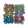
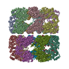
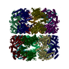
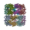
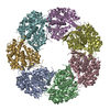
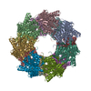
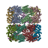
| Title | C7 Symmetrized Structure of Unliganded GroEL at 4.7 Angstrom Resolution from CryoEM | ||||||
 Components Components | 60 kDa chaperonin | ||||||
 Keywords Keywords |  CHAPERONE / GroEL / ATP-binding / Cell cycle / Cell division / Nucleotide-binding / Phosphoprotein CHAPERONE / GroEL / ATP-binding / Cell cycle / Cell division / Nucleotide-binding / Phosphoprotein | ||||||
| Function / homology |  Function and homology information Function and homology informationGroEL-GroES complex / chaperonin ATPase / virion assembly / chaperone cofactor-dependent protein refolding / isomerase activity / ATP-dependent protein folding chaperone / response to radiation / unfolded protein binding / protein folding / response to heat ...GroEL-GroES complex / chaperonin ATPase / virion assembly / chaperone cofactor-dependent protein refolding / isomerase activity / ATP-dependent protein folding chaperone / response to radiation / unfolded protein binding / protein folding / response to heat / protein refolding / magnesium ion binding / ATP hydrolysis activity / ATP binding / membrane / identical protein binding / cytosolSimilarity search - Function | ||||||
| Biological species |  Escherichia coli (E. coli) Escherichia coli (E. coli) | ||||||
| Method | ELECTRON MICROSCOPY / single particle reconstruction / cryo EM / Resolution: 4.7 Å | ||||||
 Authors Authors | Ludtke, S.J. / Baker, M.L. / Chen, D.H. / Song, J.L. / Chuang, D. / Chiu, W. | ||||||
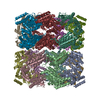
 Citation Citation | Journal: Structure / Year: 2008 Title: De novo backbone trace of GroEL from single particle electron cryomicroscopy. Authors: Steven J Ludtke / Matthew L Baker / Dong-Hua Chen / Jiu-Li Song / David T Chuang / Wah Chiu /  Abstract: In this work, we employ single-particle electron cryo-microscopy (cryo-EM) to reconstruct GroEL to approximately 4 A resolution with both D7 and C7 symmetry. Using a newly developed skeletonization ...In this work, we employ single-particle electron cryo-microscopy (cryo-EM) to reconstruct GroEL to approximately 4 A resolution with both D7 and C7 symmetry. Using a newly developed skeletonization algorithm and secondary structure element identification in combination with sequence-based secondary structure prediction, we demonstrate that it is possible to achieve a de novo Calpha trace directly from a cryo-EM reconstruction. The topology of our backbone trace is completely accurate, though subtle alterations illustrate significant differences from existing crystal structures. In the map with C7 symmetry, the seven monomers in each ring are identical; however, the subunits have a subtly different structure in each ring, particularly in the equatorial domain. These differences include an asymmetric salt bridge, density in the nucleotide-binding pocket of only one ring, and small shifts in alpha helix positions. This asymmetric conformation is different from previous asymmetric structures, including GroES-bound GroEL, and may represent a "primed state" in the chaperonin pathway. | ||||||
| History |
|
- Structure visualization
| Movie |
Movie viewer |
|---|---|
| Structure viewer | Molecule: MolmilJmol/JSmol |
- Downloads & links
-Download
| PDBx/mmCIF format | 3c9v.cif.gz | 192.3 KB | Display | PDBx/mmCIF format |
|---|---|---|---|---|
| PDB format | pdb3c9v.ent.gz | 128.5 KB | Display | PDB format |
| PDBx/mmJSON format | 3c9v.json.gz | Tree view | PDBx/mmJSON format | |
| Others |  Other downloads Other downloads |
-Validation report
| Arichive directory | https://data.pdbj.org/pub/pdb/validation_reports/c9/3c9vftp://data.pdbj.org/pub/pdb/validation_reports/c9/3c9v | HTTPS FTP |
|---|
-Related structure data
| Related structure data | 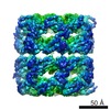 5002MC 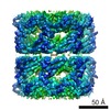 5001C 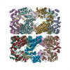 3cauC M: map data used to model this data C: citing same article ( |
|---|---|
| Similar structure data |
-Links
 PDBj
PDBj
- Assembly
| Deposited unit | 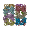
|
|---|---|
| 1 |
|
-Components
| #1: Protein | Mass: 55463.387 Da / Num. of mol.: 14 / Fragment: residues 2-527 Source method: isolated from a genetically manipulated source Source: (gene. exp.) Escherichia coli (E. coli) / Gene: groL, groEL, mopA / Plasmid: pGroESL / Production host: Escherichia coli (E. coli) / Strain (production host): ESts CG-712 / References: UniProt: P0A6F5 |
|---|
-Experimental details
-Experiment
| Experiment | Method: ELECTRON MICROSCOPY |
|---|---|
| EM experiment | Aggregation state: PARTICLE / 3D reconstruction method: single particle reconstruction |
- Sample preparation
| Component | Name: GroEL / Type: COMPLEX / Details: 14-mer. Two back to back homo-heptameric rings. |
|---|---|
| Buffer solution | Name: 20 mM Tris.HCl, pH 7.5, 50 mM MgCl2 / pH: 7.5 / Details: 20 mM Tris.HCl, pH 7.5, 50 mM MgCl2 |
| Specimen | Embedding applied: NO / Shadowing applied: NO / Staining applied: NO / Vitrification applied: YES |
| Vitrification | Instrument: FEI VITROBOT MARK I / Cryogen name: ETHANE / Details: ETHANE. Vitrobot, blot for 2 sec. |
- Electron microscopy imaging
| Microscopy | Model: JEOL 3200FS / Date: Jan 1, 2005 |
|---|---|
| Electron gun | Electron source: FIELD EMISSION GUN / Accelerating voltage: 300 kV / Illumination mode: FLOOD BEAM |
| Electron lens | Mode: BRIGHT FIELDBright-field microscopy / Nominal magnification: 60000 X / Nominal defocus max: 2300 nm / Nominal defocus min: 900 nm / Cs: 1.6 mm |
| Specimen holder | Temperature: 4 K / Tilt angle max: 0 ° / Tilt angle min: 0 ° |
| Image recording | Electron dose: 36 e/Å2 / Film or detector model: KODAK SO-163 FILM |
- Processing
| CTF correction | Details: per micrograph | ||||||||||||
|---|---|---|---|---|---|---|---|---|---|---|---|---|---|
| Symmetry | Point symmetry: C7 (7 fold cyclic) | ||||||||||||
| 3D reconstruction | Method: EMAN, single particle / Resolution: 4.7 Å / Num. of particles: 20401 / Nominal pixel size: 1.06 Å / Details: C7 symmetry, This entry contains CA atom only / Symmetry type: POINT | ||||||||||||
| Refinement step | Cycle: LAST
|
