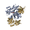
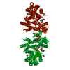
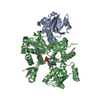
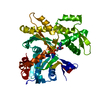
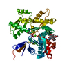
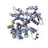
SHEET THE SHEET STRUCTURE OF THIS MOLECULE IS BIFURCATED. IN ORDER TO REPRESENT THIS FEATURE IN ... SHEET THE SHEET STRUCTURE OF THIS MOLECULE IS BIFURCATED. IN ORDER TO REPRESENT THIS FEATURE IN THE SHEET RECORDS BELOW, TWO SHEETS ARE DEFINED.
Resolution: 3.2→20 Å / Isotropic thermal model: GROUP / Cross valid method: THROUGHOUT / σ(F): 3 / Stereochemistry target values: TWIN_LSQ Details: 1. BULK SOLVENT MODEL USED. 2. THE DATA WERE MEROHEDRALLY TWINNED IN THE SPACE GROUP P61 WITH THE TWIN LAW H,-H-K,-L. THE TWINNING FRACTION WAS 0.432. 3. THE REFINEMENT WAS AGAINST THE NON- ...Details: 1. BULK SOLVENT MODEL USED. 2. THE DATA WERE MEROHEDRALLY TWINNED IN THE SPACE GROUP P61 WITH THE TWIN LAW H,-H-K,-L. THE TWINNING FRACTION WAS 0.432. 3. THE REFINEMENT WAS AGAINST THE NON-DETWINNED DATA USING CNS IN THE TWIN MODE. 4. ALL R FACTORS, ARE SO CALLED TWINNED R FACTORS, AND ARE CALCULATED FROM THE DIFFERENCE BETWEEN THE TWINNED FOBS AND THE TWINNED FCALC. 5. STRICT NCS BETWEEN MOLECULES A, D,F AND B,E,G WERE FIRST USED. RESTRAINT NCS WERE USED FOR THE LATEST STAGES OF REFINEMENT.
In the structure databanks used in Yorodumi, some data are registered as the other names, "COVID-19 virus" and "2019-nCoV". Here are the details of the virus and the list of structure data.
Jan 31, 2019. EMDB accession codes are about to change! (news from PDBe EMDB page)
EMDB accession codes are about to change! (news from PDBe EMDB page)
The allocation of 4 digits for EMDB accession codes will soon come to an end. Whilst these codes will remain in use, new EMDB accession codes will include an additional digit and will expand incrementally as the available range of codes is exhausted. The current 4-digit format prefixed with “EMD-” (i.e. EMD-XXXX) will advance to a 5-digit format (i.e. EMD-XXXXX), and so on. It is currently estimated that the 4-digit codes will be depleted around Spring 2019, at which point the 5-digit format will come into force.
The EM Navigator/Yorodumi systems omit the EMD- prefix.
Related info.:Q: What is EMD? / ID/Accession-code notation in Yorodumi/EM Navigator
Yorodumi is a browser for structure data from EMDB, PDB, SASBDB, etc.
This page is also the successor to EM Navigator detail page, and also detail information page/front-end page for Omokage search.
The word "yorodu" (or yorozu) is an old Japanese word meaning "ten thousand". "mi" (miru) is to see.
Related info.:EMDB / PDB / SASBDB / Comparison of 3 databanks / Yorodumi Search / Aug 31, 2016. New EM Navigator & Yorodumi / Yorodumi Papers / Jmol/JSmol / Function and homology information / Changes in new EM Navigator and Yorodumi
 Movie
Movie Controller

 Yorodumi
 Open data
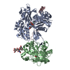
 Basic information
 Components
 Keywords
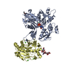
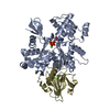
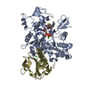
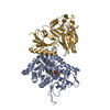
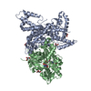
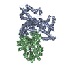
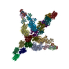
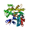
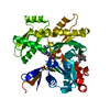
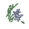
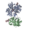
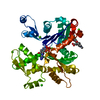
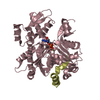
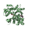
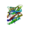
 STRUCTURAL PROTEIN / ACTIN-BINDING /
  Function and homology information

 Authors
 Citation
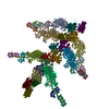
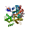
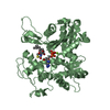
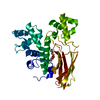
 Structure visualization
 Downloads & links
 Other downloads
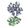


 PDBj
PDBj


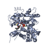
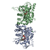
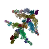
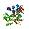
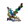
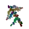
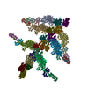
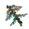
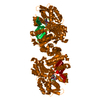
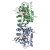
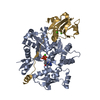
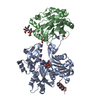
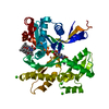
 Assembly
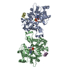


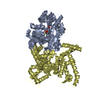
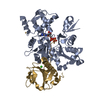
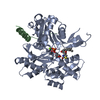
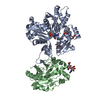
 Mass: 507.181 Da / Num. of mol.: 2 / Source method: obtained synthetically / Formula: C10H16N5O13P3 / Comment: ATP, energy-carrying molecule*YM
 Mass: 40.078 Da / Num. of mol.: 2 / Source method: obtained synthetically / Formula: Ca
 Sample preparation
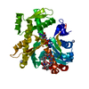
 / Beamline: ID14-4 / Wavelength: 0.97551
  Processing