 Movie
Movie Controller


+ Open data

- Basic information
| Entry | Database: PDB / ID: 2v6l | ||||||
|---|---|---|---|---|---|---|---|
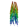
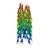
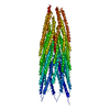
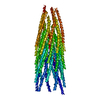
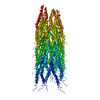
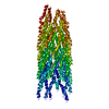
| Title | Molecular Model of a Type III Secretion System Needle | ||||||
 Components Components | MXIH | ||||||
 Keywords Keywords |  PROTEIN TRANSPORT / T3SS / VIRULENCE / TRANSPORT PROTEIN TRANSPORT / T3SS / VIRULENCE / TRANSPORT | ||||||
| Function / homology |  Function and homology informationtype III protein secretion system complex / protein secretion by the type III secretion system / cell surface / extracellular region / identical protein binding Function and homology informationtype III protein secretion system complex / protein secretion by the type III secretion system / cell surface / extracellular region / identical protein bindingSimilarity search - Function | ||||||
| Biological species |  SHIGELLA FLEXNERI (bacteria) SHIGELLA FLEXNERI (bacteria) | ||||||
| Method | ELECTRON MICROSCOPY / helical reconstruction / negative staining / Resolution: 16 Å | ||||||
| Model type details | CA ATOMS ONLY, CHAIN A, B, C, D, E, F, G, H, I, J, K, L, M, N, O, P, Q, R, S, T, U, V, W, X, Y, Z, 1, 0 | ||||||
 Authors Authors | Deane, J.E. / Roversi, P. / Cordes, F.S. / Johnson, S. / Kenjale, R. / Daniell, S. / Booy, F. / Picking, W.L. / Picking, W.D. / Blocker, A.J. / Lea, S.M. | ||||||
 Citation Citation | Journal: Proc Natl Acad Sci U S A / Year: 2006 Title: Molecular model of a type III secretion system needle: Implications for host-cell sensing. Authors: Janet E Deane / Pietro Roversi / Frank S Cordes / Steven Johnson / Roma Kenjale / Sarah Daniell / Frank Booy / William D Picking / Wendy L Picking / Ariel J Blocker / Susan M Lea /  Abstract: Type III secretion systems are essential virulence determinants for many Gram-negative bacterial pathogens. The type III secretion system consists of cytoplasmic, transmembrane, and extracellular ...Type III secretion systems are essential virulence determinants for many Gram-negative bacterial pathogens. The type III secretion system consists of cytoplasmic, transmembrane, and extracellular domains. The extracellular domain is a hollow needle protruding above the bacterial surface and is held within a basal body that traverses both bacterial membranes. Effector proteins are translocated, via this external needle, directly into host cells, where they subvert normal cell functions to aid infection. Physical contact with host cells initiates secretion and leads to formation of a pore, thought to be contiguous with the needle channel, in the host-cell membrane. Here, we report the crystal structure of the Shigella flexneri needle subunit MxiH and a complete model for the needle assembly built into our three-dimensional EM reconstruction. The model, combined with mutagenesis data, reveals that signaling of host-cell contact is relayed through the needle via intersubunit contacts and suggests a mode of binding for a tip complex. #1: Journal: J Biol Chem / Year: 2003Title: Helical structure of the needle of the type III secretion system of Shigella flexneri. Authors: Frank S Cordes / Kaoru Komoriya / Eric Larquet / Shixin Yang / Edward H Egelman / Ariel Blocker / Susan M Lea / Abstract: Gram-negative bacteria commonly interact with animal and plant hosts using type III secretion systems (TTSSs) for translocation of proteins into eukaryotic cells during infection. 10 of the 25 TTSS- ...Gram-negative bacteria commonly interact with animal and plant hosts using type III secretion systems (TTSSs) for translocation of proteins into eukaryotic cells during infection. 10 of the 25 TTSS-encoding genes are homologous to components of the bacterial flagellar basal body, which the TTSS needle complex morphologically resembles. This indicates a common ancestry, although no TTSS sequence homologues for the genes encoding the flagellum are found. We here present an approximately 16-A structure of the central component, the needle, of the TTSS. Although the needle subunit is significantly smaller and shares no sequence homology with the flagellar hook and filament, it shares a common helical architecture ( approximately 5.6 subunits/turn, 24-A helical pitch). This common architecture implies that there will be further mechanistic analogies in the functioning of these two bacterial systems. | ||||||
| History |
|
- Structure visualization
| Movie |
Movie viewer |
|---|---|
| Structure viewer | Molecule: MolmilJmol/JSmol |
- Downloads & links
-Download
| PDBx/mmCIF format | 2v6l.cif.gz | 62.9 KB | Display | PDBx/mmCIF format |
|---|---|---|---|---|
| PDB format | pdb2v6l.ent.gz | 46.2 KB | Display | PDB format |
| PDBx/mmJSON format | 2v6l.json.gz | Tree view | PDBx/mmJSON format | |
| Others |  Other downloads Other downloads |
-Validation report
| Arichive directory | https://data.pdbj.org/pub/pdb/validation_reports/v6/2v6lftp://data.pdbj.org/pub/pdb/validation_reports/v6/2v6l | HTTPS FTP |
|---|
-Related structure data
| Related structure data | 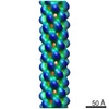 1416M  2ca5C C: citing same article ( M: map data used to model this data |
|---|---|
| Similar structure data |
-Links
 PDBj
PDBj- Assembly
| Deposited unit | 
|
|---|---|
| 1 |
|
-Components
| #1: Protein | Mass: 9272.265 Da / Num. of mol.: 28 Source method: isolated from a genetically manipulated source Details: RESIDUES 2-19 WERE MODELLED AS AN ALPHA HELIX. RESIDUES 20-80 COME FROM MOLECULE A OF PDB ENTRY 2CA5 RESIDUES 81-85 WERE MODELLED AS AN ALPHA HELIX. Source: (gene. exp.) SHIGELLA FLEXNERI (bacteria) / Strain: PWR100 / Plasmid: PET22B / Production host: Escherichia coli BL21(DE3) (bacteria) / References: UniProt: P0A223 |
|---|
-Experimental details
-Experiment
| Experiment | Method: ELECTRON MICROSCOPY |
|---|---|
| EM experiment | Aggregation state: FILAMENT / 3D reconstruction method: helical reconstruction |
- Sample preparation
| Component | Name: SHIGELLA FLEXNERI TYPE THREE SECRETION NEEDLE / Type: COMPLEX / Details: STAINED WITH A DROP OF 2% URANYL ACETATE, PH 7.5 |
|---|---|
| Buffer solution | Name: 10 MM TRIS / pH: 7.5 / Details: 10 MM TRIS |
| Specimen | Conc.: 0.03 mg/ml / Embedding applied: NO / Shadowing applied: NO / Staining applied: YES / Vitrification applied: NO |
| EM staining | Type: NEGATIVE / Material: Uranyl Acetate |
| Vitrification | Cryogen name: ETHANE |
- Electron microscopy imaging
| Microscopy | Model: FEI/PHILIPS CM200T / Details: OTHER |
|---|---|
| Electron gun | Electron source: LAB6 / Accelerating voltage: 120 kV / Illumination mode: FLOOD BEAM |
| Electron lens | Mode: BRIGHT FIELDBright-field microscopy / Nominal magnification: 60000 X / Nominal defocus max: 700 nm / Nominal defocus min: 700 nm |
| Image recording | Electron dose: 10 e/Å2 / Film or detector model: GENERIC FILM |
| Radiation wavelength | Relative weight: 1 |
- Processing
| EM software |
| ||||||||||||
|---|---|---|---|---|---|---|---|---|---|---|---|---|---|
| CTF correction | Details: GRID | ||||||||||||
| 3D reconstruction | Method: RECIPROCAL SPACE RIGID BODY FIT / Resolution: 16 Å / Num. of particles: 5000 / Nominal pixel size: 2.65 Å Details: A SET OF X-RAY STRUCTURE FACTORS WERE COMPUTED BY PLACING THE EM DENSITY IN A CRYSTAL CELL. THE MXIH MONOMER WAS RIGID BODY REFINED AGAINST THESE DATA WITH HARD NCS CONSTRAINTS. THE EM ...Details: A SET OF X-RAY STRUCTURE FACTORS WERE COMPUTED BY PLACING THE EM DENSITY IN A CRYSTAL CELL. THE MXIH MONOMER WAS RIGID BODY REFINED AGAINST THESE DATA WITH HARD NCS CONSTRAINTS. THE EM DENSITY FOR THE NEEDLE WAS PUT IN THIS P1 CRYSTAL CELL SO THAT SINGLE CRYSTAL X-RAY MODEL REFINEMENT PROGRAMS COULD BE USED TO FIT THE MODEL. Symmetry type: HELICAL | ||||||||||||
| Atomic model building | Protocol: OTHER / Space: RECIPROCAL / Target criteria: Cross-correlation coefficient Details: METHOD--RECIPROCAL SPACE FIT REFINEMENT PROTOCOL--X-RAY | ||||||||||||
| Atomic model building | PDB-ID: 2CA5 | ||||||||||||
| Refinement | Highest resolution: 16 Å | ||||||||||||
| Refinement step | Cycle: LAST / Highest resolution: 16 Å
|
