 Movie
Movie Controller

[English] 日本語
 Yorodumi
- PDB-2ff6: Crystal structure of Gelsolin domain 1:ciboulot domain 2 hybrid i... -
+ Open data

- Basic information
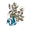
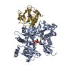
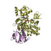
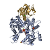
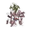
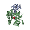
| Entry | Database: PDB / ID: 2ff6 | ||||||
|---|---|---|---|---|---|---|---|
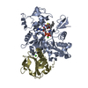
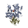
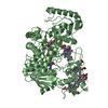
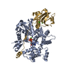
| Title | Crystal structure of Gelsolin domain 1:ciboulot domain 2 hybrid in complex with actin | ||||||
 Components Components |
| ||||||
 Keywords Keywords |  STRUCTURAL PROTEIN/CONTRACTILE PROTEIN / Protein-Protein complex / STRUCTURAL PROTEIN-CONTRACTILE PROTEIN COMPLEX STRUCTURAL PROTEIN/CONTRACTILE PROTEIN / Protein-Protein complex / STRUCTURAL PROTEIN-CONTRACTILE PROTEIN COMPLEX | ||||||
| Function / homology |  Function and homology information Function and homology informationlarval central nervous system remodeling / striated muscle atrophy / regulation of establishment of T cell polarity / regulation of plasma membrane raft polarization / regulation of receptor clustering / renal protein absorption / positive regulation of keratinocyte apoptotic process / positive regulation of protein processing in phagocytic vesicle / positive regulation of actin nucleation / phosphatidylinositol 3-kinase catalytic subunit binding ...larval central nervous system remodeling / striated muscle atrophy / regulation of establishment of T cell polarity / regulation of plasma membrane raft polarization / regulation of receptor clustering / renal protein absorption / positive regulation of keratinocyte apoptotic process / positive regulation of protein processing in phagocytic vesicle / positive regulation of actin nucleation / phosphatidylinositol 3-kinase catalytic subunit binding / positive regulation of cysteine-type endopeptidase activity involved in apoptotic signaling pathway / actin cap / sequestering of actin monomers / regulation of podosome assembly / myosin II binding / negative regulation of viral entry into host cell / actin filament severing / actin filament capping / barbed-end actin filament capping / actin filament depolymerization / actin polymerization or depolymerization / cell projection assembly / cardiac muscle cell contraction / podosome / sarcoplasm / Sensory processing of sound by outer hair cells of the cochlea / relaxation of cardiac muscle / cytoskeletal motor activator activity / phagocytosis, engulfment / cortical actin cytoskeleton / tropomyosin binding / myosin heavy chain binding / mesenchyme migration / troponin I binding / hepatocyte apoptotic process / actin filament bundle / filamentous actin / actin filament bundle assembly / skeletal muscle thin filament assembly / striated muscle thin filament / skeletal muscle myofibril / cilium assembly / actin monomer binding / Caspase-mediated cleavage of cytoskeletal proteins / skeletal muscle fiber development / phagocytic vesicle / stress fiber / titin binding / phosphatidylinositol-4,5-bisphosphate binding / response to muscle stretch / actin filament polymerization / filopodium / central nervous system development / actin filament organization / actin filament / Hydrolases; Acting on acid anhydrides; Acting on acid anhydrides to facilitate cellular and subcellular movement / brain development / protein destabilization / cellular response to type II interferon / calcium-dependent protein binding / actin filament binding / actin cytoskeleton / lamellipodium / cell body / actin binding / blood microparticle / secretory granule lumen / ficolin-1-rich granule lumen / amyloid fibril formation / cytoskeleton / hydrolase activity / Amyloid fiber formation / protein domain specific binding / focal adhesion / calcium ion binding / Neutrophil degranulation / positive regulation of gene expression / magnesium ion binding / extracellular space / extracellular exosome / extracellular region / ATP binding / identical protein binding / plasma membrane / cytosol / cytoplasmSimilarity search - Function | ||||||
| Biological species |  Homo sapiens (human) Homo sapiens (human) Drosophila melanogaster (fruit fly) Drosophila melanogaster (fruit fly) Oryctolagus cuniculus (rabbit) Oryctolagus cuniculus (rabbit) | ||||||
| Method | X-RAY DIFFRACTION / SYNCHROTRON / MOLECULAR REPLACEMENT / Resolution: 2.05 Å | ||||||
 Authors Authors | Aguda, A.H. / Xue, B. / Robinson, R.C. | ||||||
 Citation Citation | Journal: Structure / Year: 2006 Title: The Structural Basis of Actin Interaction with Multiple WH2/beta-Thymosin Motif-Containing Proteins Authors: Aguda, A.H. / Xue, B. / Irobi, E. / Preat, T. / Robinson, R.C. | ||||||
| History |
|
- Structure visualization
| Structure viewer | Molecule: MolmilJmol/JSmol |
|---|
- Downloads & links
-Download
| PDBx/mmCIF format | 2ff6.cif.gz | 116.5 KB | Display | PDBx/mmCIF format |
|---|---|---|---|---|
| PDB format | pdb2ff6.ent.gz | 91.9 KB | Display | PDB format |
| PDBx/mmJSON format | 2ff6.json.gz | Tree view | PDBx/mmJSON format | |
| Others |  Other downloads Other downloads |
-Validation report
| Arichive directory | https://data.pdbj.org/pub/pdb/validation_reports/ff/2ff6ftp://data.pdbj.org/pub/pdb/validation_reports/ff/2ff6 | HTTPS FTP |
|---|
-Related structure data


-Links
 PDBj
PDBj


- Assembly
| Deposited unit | 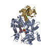
| ||||||||
|---|---|---|---|---|---|---|---|---|---|
| 1 |
| ||||||||
| Unit cell |
|
-Components
-Protein , 2 types, 2 molecules GA
| #1: Protein | / Actin-depolymerizing factor / ADF / Brevin / AGEL Mass: 16867.998 Da / Num. of mol.: 1 / Fragment: Gelsolin domain 1 Source method: isolated from a genetically manipulated source Source: (gene. exp.) Homo sapiens (human) / Plasmid: pHis8-3 / Production host:  Escherichia coli (E. coli) / References: UniProt: P06396 Escherichia coli (E. coli) / References: UniProt: P06396 |
|---|---|
| #3: Protein | / Alpha-actin-1 Mass: 41862.613 Da / Num. of mol.: 1 / Source method: isolated from a natural source / Source: (natural) Oryctolagus cuniculus (rabbit) / Tissue: skeletal muscle / References: UniProt: P68135 |
-Protein/peptide , 1 types, 1 molecules H
| #2: Protein/peptide | Mass: 3093.301 Da / Num. of mol.: 1 / Fragment: ciboulot domain 2 Source method: isolated from a genetically manipulated source Source: (gene. exp.) Drosophila melanogaster (fruit fly) / Plasmid: pHis8-3 / Production host: Escherichia coli (E. coli) / References: UniProt: Q8IRS7 |
|---|
-Non-polymers , 3 types, 281 molecules 


| #4: Chemical |  Mass: 40.078 Da / Num. of mol.: 3 / Source method: obtained synthetically / Formula: Ca Mass: 40.078 Da / Num. of mol.: 3 / Source method: obtained synthetically / Formula: Ca#5: Chemical | ChemComp-ATP / | Adenosine triphosphate Mass: 507.181 Da / Num. of mol.: 1 / Source method: obtained synthetically / Formula: C10H16N5O13P3 / Comment: ATP, energy-carrying molecule*YM Mass: 507.181 Da / Num. of mol.: 1 / Source method: obtained synthetically / Formula: C10H16N5O13P3 / Comment: ATP, energy-carrying molecule*YM#6: Water | ChemComp-HOH / | WaterMass: 18.015 Da / Num. of mol.: 277 / Source method: isolated from a natural source / Formula: H2O |
|---|
-Experimental details
-Experiment
| Experiment | Method: X-RAY DIFFRACTION / Number of used crystals: 1 |
|---|
- Sample preparation
| Crystal | Density Matthews: 2.6 Å3/Da / Density % sol: 52.76 % |
|---|---|
| Crystal grow | Temperature: 293.15 K / Method: vapor diffusion, sitting drop / pH: 7 Details: 7% PEG 3000, 0.1M Hepes, pH 7.0, VAPOR DIFFUSION, SITTING DROP, temperature 293.15K |
-Data collection
| Diffraction | Mean temperature: 100 K |
|---|---|
| Diffraction source | Source: SYNCHROTRON / Site: MAX II  / Beamline: I711 / Wavelength: 0.934 Å / Beamline: I711 / Wavelength: 0.934 Å |
| Detector | Type: MARRESEARCH / Detector: IMAGE PLATE / Date: Oct 16, 2004 |
| Radiation | Monochromator: Si(111) / Protocol: SINGLE WAVELENGTH / Monochromatic (M) / Laue (L): M / Scattering type: x-ray |
| Radiation wavelength | Wavelength: 0.934 Å / Relative weight: 1 |
| Reflection | Resolution: 2.05→79.31 Å / Num. all: 38456 / Num. obs: 36145 / % possible obs: 99.04 % / Observed criterion σ(F): 0 / Observed criterion σ(I): 0 |
| Reflection shell | Resolution: 2.05→2.1 Å / % possible all: 99.2 |
- Processing
| Software |
| |||||||||||||||||||||||||||||||||||||||||||||||||||||||||||||||||||||||||||||||||||||||||||||||
|---|---|---|---|---|---|---|---|---|---|---|---|---|---|---|---|---|---|---|---|---|---|---|---|---|---|---|---|---|---|---|---|---|---|---|---|---|---|---|---|---|---|---|---|---|---|---|---|---|---|---|---|---|---|---|---|---|---|---|---|---|---|---|---|---|---|---|---|---|---|---|---|---|---|---|---|---|---|---|---|---|---|---|---|---|---|---|---|---|---|---|---|---|---|---|---|---|
| Refinement | Method to determine structure: MOLECULAR REPLACEMENT / Resolution: 2.05→29.5 Å / Cor.coef. Fo:Fc: 0.937 / Cor.coef. Fo:Fc free: 0.912 / SU B: 4.355 / SU ML: 0.119 / Cross valid method: THROUGHOUT / σ(F): 0 / ESU R: 0.198 / ESU R Free: 0.169 / Stereochemistry target values: MAXIMUM LIKELIHOOD / Details: HYDROGENS HAVE BEEN ADDED IN THE RIDING POSITIONS
| |||||||||||||||||||||||||||||||||||||||||||||||||||||||||||||||||||||||||||||||||||||||||||||||
| Solvent computation | Ion probe radii: 0.8 Å / Shrinkage radii: 0.8 Å / VDW probe radii: 1.2 Å / Solvent model: MASK | |||||||||||||||||||||||||||||||||||||||||||||||||||||||||||||||||||||||||||||||||||||||||||||||
| Displacement parameters | Biso mean: 23.681 Å2
| |||||||||||||||||||||||||||||||||||||||||||||||||||||||||||||||||||||||||||||||||||||||||||||||
| Refinement step | Cycle: LAST / Resolution: 2.05→29.5 Å
| |||||||||||||||||||||||||||||||||||||||||||||||||||||||||||||||||||||||||||||||||||||||||||||||
| Refine LS restraints |
| |||||||||||||||||||||||||||||||||||||||||||||||||||||||||||||||||||||||||||||||||||||||||||||||
| LS refinement shell | Resolution: 2.05→2.103 Å / Total num. of bins used: 20
|
