[English] 日本語
Yorodumi
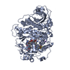
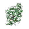
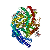
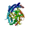
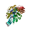
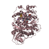
- PDB-2bf1: Structure of an unliganded and fully-glycosylated SIV gp120 envel... -

+
Open data


ID or keywords:

Loading...

-
Basic information

Entry
Database: PDB / ID: 2bf1
TitleStructure of an unliganded and fully-glycosylated SIV gp120 envelope glycoprotein
ComponentsEXTERIOR MEMBRANE GLYCOPROTEIN GP120
KeywordsVIRUS PROTEIN / SIV / GP120 / ENVELOPE GLYCOPROTEIN / AIDS / COAT PROTEIN
Function / homology
Function and homology information


membrane fusion involved in viral entry into host cell / host cell endosome membrane / symbiont entry into host cell / viral envelope / structural molecule activity / virion attachment to host cell / host cell plasma membrane / virion membrane / plasma membrane
Similarity search - Function
HIV Envelope Protein Gp120; Chain G / Human immunodeficiency virus 1, Gp160, envelope glycoprotein / Retroviral envelope protein / Retroviral envelope protein GP41-like / Gp120 core superfamily / Envelope glycoprotein GP120 / Human immunodeficiency virus 1, envelope glycoprotein Gp120 / Beta Complex / Mainly Beta
Similarity search - Domain/homology
Envelope glycoprotein gp160
Similarity search - Component
Biological speciesSIMIAN IMMUNODEFICIENCY VIRUS
MethodX-RAY DIFFRACTION / SYNCHROTRON / MIRAS / Resolution: 4 Å
AuthorsChen, B. / Vogan, E.M. / Gong, H. / Skehel, J.J. / Wiley, D.C. / Harrison, S.C.
Citation
Journal: Nature / Year: 2005
Title: Structure of an Unliganded Simian Immunodeficiency Virus Gp120 Core
Authors: Chen, B. / Vogan, E.M. / Gong, H. / Skehel, J.J. / Wiley, D.C. / Harrison, S.C.
#1: Journal: Structure / Year: 2005
Title: Determining the Structure of the Unliganded and Fully-Glycosylated Siv Gp120 Envelope Glycoprotein.
Authors: Chen, B. / Vogan, E.M. / Gong, H. / Skehel, J.J. / Wiley, D.C. / Harrison, S.C.
History
DepositionDec 2, 2004Deposition site: PDBE / Processing site: PDBE
Revision 1.0Feb 17, 2005Provider: repository / Type: Initial release
Revision 1.1May 8, 2011Group: Version format compliance
Revision 1.2Jul 13, 2011Group: Version format compliance
Revision 2.0Aug 29, 2018Group: Advisory / Atomic model ...Advisory / Atomic model / Data collection / Derived calculations / Non-polymer description / Structure summary
Category: atom_site / chem_comp ...atom_site / chem_comp / database_PDB_caveat / entity / pdbx_entity_nonpoly / pdbx_nonpoly_scheme / pdbx_validate_chiral / struct_asym / struct_conn / struct_site_gen
Item: _chem_comp.formula / _chem_comp.formula_weight ..._chem_comp.formula / _chem_comp.formula_weight / _chem_comp.id / _chem_comp.mon_nstd_flag / _chem_comp.name / _chem_comp.type / _pdbx_nonpoly_scheme.entity_id / _pdbx_nonpoly_scheme.mon_id / _pdbx_nonpoly_scheme.pdb_mon_id / _struct_asym.entity_id / _struct_conn.ptnr1_auth_comp_id / _struct_conn.ptnr1_label_comp_id / _struct_conn.ptnr2_auth_comp_id / _struct_conn.ptnr2_label_comp_id / _struct_site_gen.auth_comp_id / _struct_site_gen.label_comp_id
Revision 2.1Apr 3, 2019Group: Data collection / Derived calculations / Source and taxonomy
Category: entity_src_gen / struct_conn
Item: _entity_src_gen.pdbx_host_org_cell_line / _struct_conn.pdbx_leaving_atom_flag
Revision 2.2May 8, 2019Group: Data collection / Experimental preparation / Category: exptl_crystal_grow / Item: _exptl_crystal_grow.temp
Revision 3.0Jul 29, 2020Group: Advisory / Atomic model ...Advisory / Atomic model / Data collection / Derived calculations / Other / Structure summary
Category: atom_site / chem_comp ...atom_site / chem_comp / database_PDB_caveat / entity / pdbx_branch_scheme / pdbx_chem_comp_identifier / pdbx_database_status / pdbx_entity_branch / pdbx_entity_branch_descriptor / pdbx_entity_branch_link / pdbx_entity_branch_list / pdbx_entity_nonpoly / pdbx_nonpoly_scheme / pdbx_struct_assembly_gen / pdbx_validate_chiral / pdbx_validate_close_contact / pdbx_validate_symm_contact / struct_asym / struct_conn / struct_site / struct_site_gen
Item: _atom_site.B_iso_or_equiv / _atom_site.Cartn_x ..._atom_site.B_iso_or_equiv / _atom_site.Cartn_x / _atom_site.Cartn_y / _atom_site.Cartn_z / _atom_site.auth_asym_id / _atom_site.auth_atom_id / _atom_site.auth_comp_id / _atom_site.auth_seq_id / _atom_site.label_asym_id / _atom_site.label_atom_id / _atom_site.label_comp_id / _atom_site.label_entity_id / _atom_site.type_symbol / _chem_comp.name / _chem_comp.type / _database_PDB_caveat.text / _pdbx_database_status.status_code_sf / _pdbx_struct_assembly_gen.asym_id_list / _pdbx_validate_chiral.auth_asym_id / _pdbx_validate_chiral.auth_seq_id / _pdbx_validate_close_contact.auth_asym_id_2 / _pdbx_validate_close_contact.auth_seq_id_2 / _pdbx_validate_symm_contact.auth_asym_id_1 / _pdbx_validate_symm_contact.auth_asym_id_2 / _pdbx_validate_symm_contact.auth_seq_id_1 / _pdbx_validate_symm_contact.auth_seq_id_2 / _struct_conn.pdbx_dist_value / _struct_conn.pdbx_leaving_atom_flag / _struct_conn.pdbx_role / _struct_conn.ptnr1_auth_asym_id / _struct_conn.ptnr1_auth_seq_id / _struct_conn.ptnr1_label_asym_id / _struct_conn.ptnr1_label_atom_id / _struct_conn.ptnr2_auth_asym_id / _struct_conn.ptnr2_auth_comp_id / _struct_conn.ptnr2_auth_seq_id / _struct_conn.ptnr2_label_asym_id / _struct_conn.ptnr2_label_comp_id
Description: Carbohydrate remediation / Provider: repository / Type: Remediation

-
Structure visualization

Structure viewerMolecule:
MolmilJmol/JSmol

Downloads & links

-
Assembly

Deposited unit
A: EXTERIOR MEMBRANE GLYCOPROTEIN GP120
hetero molecules


Theoretical massNumber of molelcules
Total (without water)46,04814
Polymers36,8341
Non-polymers9,21413
Water0
1


  • Idetical with deposited unit
  • defined by author&software
TypeNameSymmetry operationNumber
identity operation1_555x,y,z1
MethodPQS
Unit cell
Length a, b, c (Å)108.048, 108.048, 117.701
Angle α, β, γ (deg.)90.00, 90.00, 90.00
Int Tables number96
Space group name H-MP43212
DetailsALTHOUGH THIS ENTRY DESCRIBES THE MONOMERIC STRUCTURE OFGP120, A THEORETICAL MODEL OF THE TRIMERIC FORM OF THEPROTEIN HAS BEEN GENERATED. THE DETAILS OF THE TRIMER ANDTHE MATRICES RELATING CHAIN A OF THIS ENTRY TO THECONSTITUENTS OF THE TRIMERIC STRUCTURE CAN BE FOUND INREMARK 400 BELOW.

-
Components

-
Protein , 1 types, 1 molecules A

#1: Protein EXTERIOR MEMBRANE GLYCOPROTEIN GP120 / SIV GP120


Mass: 36833.723 Da / Num. of mol.: 1 / Fragment: GP120 CORE, RESIDUES 66-109,209-311,342-502
Source method: isolated from a genetically manipulated source
Source: (gene. exp.) SIMIAN IMMUNODEFICIENCY VIRUS / Strain: MAC32H / Plasmid: PSIVGP120CORE / Cell line (production host): High Five / Production host: TRICHOPLUSIA NI (cabbage looper) / References: UniProt: Q07374

-
Sugars , 8 types, 13 molecules

#2: Polysaccharide beta-D-mannopyranose-(1-4)-2-acetamido-2-deoxy-beta-D-glucopyranose-(1-4)-[alpha-L-fucopyranose-(1- ...beta-D-mannopyranose-(1-4)-2-acetamido-2-deoxy-beta-D-glucopyranose-(1-4)-[alpha-L-fucopyranose-(1-6)]2-acetamido-2-deoxy-beta-D-glucopyranose


Type: oligosaccharide / Mass: 732.682 Da / Num. of mol.: 3
Source method: isolated from a genetically manipulated source
DescriptorTypeProgram
DManpb1-4DGlcpNAcb1-4[LFucpa1-6]DGlcpNAcb1-Glycam Condensed SequenceGMML 1.0
WURCS=2.0/3,4,3/[a2122h-1b_1-5_2*NCC/3=O][a1122h-1b_1-5][a1221m-1a_1-5]/1-1-2-3/a4-b1_a6-d1_b4-c1WURCSPDB2Glycan 1.1.0
[]{[(4+1)][a-D-GlcpNAc]{[(4+1)][b-D-GlcpNAc]{[(4+1)][b-D-Manp]{}}[(6+1)][b-L-Fucp]{}}}LINUCSPDB-CARE
#3: Polysaccharide alpha-D-mannopyranose-(1-3)-[alpha-D-mannopyranose-(1-6)]beta-D-mannopyranose-(1-4)-2-acetamido-2- ...alpha-D-mannopyranose-(1-3)-[alpha-D-mannopyranose-(1-6)]beta-D-mannopyranose-(1-4)-2-acetamido-2-deoxy-beta-D-glucopyranose-(1-4)-[alpha-L-fucopyranose-(1-6)]2-acetamido-2-deoxy-beta-D-glucopyranose


Type: oligosaccharide / Mass: 1056.964 Da / Num. of mol.: 2
Source method: isolated from a genetically manipulated source
DescriptorTypeProgram
DManpa1-3[DManpa1-6]DManpb1-4DGlcpNAcb1-4[LFucpa1-6]DGlcpNAcb1-Glycam Condensed SequenceGMML 1.0
WURCS=2.0/4,6,5/[a2122h-1b_1-5_2*NCC/3=O][a1122h-1b_1-5][a1122h-1a_1-5][a1221m-1a_1-5]/1-1-2-3-3-4/a4-b1_a6-f1_b4-c1_c3-d1_c6-e1WURCSPDB2Glycan 1.1.0
[]{[(4+1)][b-D-GlcpNAc]{[(4+1)][b-D-GlcpNAc]{[(4+1)][b-D-Manp]{[(3+1)][a-D-Manp]{}[(6+1)][a-D-Manp]{}}}[(6+1)][b-L-Fucp]{}}}LINUCSPDB-CARE
#4: Polysaccharide 2-acetamido-2-deoxy-beta-D-glucopyranose-(1-4)-[alpha-L-fucopyranose-(1-6)]2-acetamido-2-deoxy-beta- ...2-acetamido-2-deoxy-beta-D-glucopyranose-(1-4)-[alpha-L-fucopyranose-(1-6)]2-acetamido-2-deoxy-beta-D-glucopyranose


Type: oligosaccharide / Mass: 570.542 Da / Num. of mol.: 2
Source method: isolated from a genetically manipulated source
DescriptorTypeProgram
DGlcpNAcb1-4[LFucpa1-6]DGlcpNAcb1-Glycam Condensed SequenceGMML 1.0
WURCS=2.0/2,3,2/[a2122h-1b_1-5_2*NCC/3=O][a1221m-1a_1-5]/1-1-2/a4-b1_a6-c1WURCSPDB2Glycan 1.1.0
[]{[(4+1)][a-D-GlcpNAc]{[(4+1)][b-D-GlcpNAc]{}[(6+1)][b-L-Fucp]{}}}LINUCSPDB-CARE
#5: Polysaccharide 2-acetamido-2-deoxy-beta-D-glucopyranose-(1-4)-2-acetamido-2-deoxy-beta-D-glucopyranose


Type: oligosaccharide / Mass: 424.401 Da / Num. of mol.: 1
Source method: isolated from a genetically manipulated source
DescriptorTypeProgram
DGlcpNAcb1-4DGlcpNAcb1-Glycam Condensed SequenceGMML 1.0
WURCS=2.0/1,2,1/[a2122h-1b_1-5_2*NCC/3=O]/1-1/a4-b1WURCSPDB2Glycan 1.1.0
[]{[(4+1)][a-D-GlcpNAc]{[(4+1)][b-D-GlcpNAc]{}}}LINUCSPDB-CARE
#6: Polysaccharide alpha-D-mannopyranose-(1-2)-alpha-D-mannopyranose-(1-2)-alpha-D-mannopyranose-(1-3)-[alpha-D- ...alpha-D-mannopyranose-(1-2)-alpha-D-mannopyranose-(1-2)-alpha-D-mannopyranose-(1-3)-[alpha-D-mannopyranose-(1-6)]beta-D-mannopyranose-(1-4)-2-acetamido-2-deoxy-beta-D-glucopyranose-(1-4)-2-acetamido-2-deoxy-beta-D-glucopyranose


Type: oligosaccharide / Mass: 1235.105 Da / Num. of mol.: 1
Source method: isolated from a genetically manipulated source
DescriptorTypeProgram
DManpa1-2DManpa1-2DManpa1-3[DManpa1-6]DManpb1-4DGlcpNAcb1-4DGlcpNAcb1-Glycam Condensed SequenceGMML 1.0
WURCS=2.0/3,7,6/[a2122h-1b_1-5_2*NCC/3=O][a1122h-1b_1-5][a1122h-1a_1-5]/1-1-2-3-3-3-3/a4-b1_b4-c1_c3-d1_c6-g1_d2-e1_e2-f1WURCSPDB2Glycan 1.1.0
[]{[(4+1)][b-D-GlcpNAc]{[(4+1)][b-D-GlcpNAc]{[(4+1)][b-D-Manp]{[(3+1)][a-D-Manp]{[(2+1)][a-D-Manp]{[(2+1)][a-D-Manp]{}}}[(6+1)][a-D-Manp]{}}}}}LINUCSPDB-CARE
#7: Polysaccharide alpha-D-mannopyranose-(1-3)-[alpha-D-mannopyranose-(1-6)]beta-D-mannopyranose-(1-4)-2-acetamido-2- ...alpha-D-mannopyranose-(1-3)-[alpha-D-mannopyranose-(1-6)]beta-D-mannopyranose-(1-4)-2-acetamido-2-deoxy-beta-D-glucopyranose-(1-4)-2-acetamido-2-deoxy-beta-D-glucopyranose


Type: oligosaccharide / Mass: 910.823 Da / Num. of mol.: 1
Source method: isolated from a genetically manipulated source
DescriptorTypeProgram
DManpa1-3[DManpa1-6]DManpb1-4DGlcpNAcb1-4DGlcpNAcb1-Glycam Condensed SequenceGMML 1.0
WURCS=2.0/3,5,4/[a2122h-1b_1-5_2*NCC/3=O][a1122h-1b_1-5][a1122h-1a_1-5]/1-1-2-3-3/a4-b1_b4-c1_c3-d1_c6-e1WURCSPDB2Glycan 1.1.0
[]{[(4+1)][b-D-GlcpNAc]{[(4+1)][b-D-GlcpNAc]{[(4+1)][b-D-Manp]{[(3+1)][a-D-Manp]{}[(6+1)][a-D-Manp]{}}}}}LINUCSPDB-CARE
#8: Polysaccharide alpha-D-mannopyranose-(1-6)-beta-D-mannopyranose-(1-4)-2-acetamido-2-deoxy-beta-D-glucopyranose-(1- ...alpha-D-mannopyranose-(1-6)-beta-D-mannopyranose-(1-4)-2-acetamido-2-deoxy-beta-D-glucopyranose-(1-4)-2-acetamido-2-deoxy-beta-D-glucopyranose


Type: oligosaccharide / Mass: 748.682 Da / Num. of mol.: 1
Source method: isolated from a genetically manipulated source
DescriptorTypeProgram
DManpa1-6DManpb1-4DGlcpNAcb1-4DGlcpNAcb1-Glycam Condensed SequenceGMML 1.0
WURCS=2.0/3,4,3/[a2122h-1b_1-5_2*NCC/3=O][a1122h-1b_1-5][a1122h-1a_1-5]/1-1-2-3/a4-b1_b4-c1_c6-d1WURCSPDB2Glycan 1.1.0
[]{[(4+1)][a-D-GlcpNAc]{[(4+1)][b-D-GlcpNAc]{[(4+1)][b-D-Manp]{[(6+1)][a-D-Manp]{}}}}}LINUCSPDB-CARE
#9: Sugar ChemComp-NAG / 2-acetamido-2-deoxy-beta-D-glucopyranose / N-Acetylglucosamine


Type: D-saccharide, beta linking / Mass: 221.208 Da / Num. of mol.: 2
Source method: isolated from a genetically manipulated source
Formula: C8H15NO6
IdentifierTypeProgram
DGlcpNAcbCONDENSED IUPAC CARBOHYDRATE SYMBOLGMML 1.0
N-acetyl-b-D-glucopyranosamineCOMMON NAMEGMML 1.0
b-D-GlcpNAcIUPAC CARBOHYDRATE SYMBOLPDB-CARE 1.0
GlcNAcSNFG CARBOHYDRATE SYMBOLGMML 1.0

-
Details

Compound detailsGP120 IS PRODUCED FROM A PRECURSOR PROTEIN, GP160. THE PRECURSOR ASSEMBLES INTO A TRIMER, WHICH ...GP120 IS PRODUCED FROM A PRECURSOR PROTEIN, GP160. THE PRECURSOR ASSEMBLES INTO A TRIMER, WHICH REMAINS INTACT AFTER CLEAVAGE OF GP160 TO GP120 AND GP41. A THEORETICAL MODEL OF THE POSITION AND ORIENTATION OF GP120 WITHIN THIS TRIMERIC ASSEMBLY HAS BEEN GENERATED. THE MODEL LACKS THE GP41 STRUCTURE, BUT SPACE HAS BEEN LEFT FOR GP41 AT THE BASE OF THE TRIMER, RESULTING IN GAPS BETWEEN THE GP120 CHAINS OF THE MODEL. THE TRIMER MODEL CAN BE GENERATED BY APPLYING THE FOLLOWING TRANSFORMATIONS TO THE COORDINATES OF CHAIN A OF THIS PDB ENTRY: TRANS1 1 -0.932060 0.211070 0.294480 1.43530 TRANS2 1 0.071796 -0.689070 0.721130 43.48100 TRANS3 1 0.355130 0.693270 0.627100 -12.91480 TRANS1 2 0.528220 -0.702280 0.477270 36.93710 TRANS2 2 0.771290 0.161790 -0.615570 -22.98340 TRANS3 2 0.355090 0.693270 0.627130 -12.91500 TRANS1 3 0.403860 0.491250 -0.771730 -38.37270 TRANS2 3 -0.843070 0.527350 -0.105510 -20.49850 TRANS3 3 0.355140 0.693230 0.627140 -12.91380
Sequence detailsIN THE EXPRESSION CONSTRUCT, SHORT LINKERS, GAG HAVE BEEN SUBSTITUTED FOR THE V1V2 AND V3 LOOPS. ...IN THE EXPRESSION CONSTRUCT, SHORT LINKERS, GAG HAVE BEEN SUBSTITUTED FOR THE V1V2 AND V3 LOOPS. THAT IS, RESIDUES FROM 110 TO 208 WERE REPLACED BY 3 RESIDUES GAG. RESIDUES FROM 312 TO 341 WERE REPLACED BY THREE RESIDUES GAG. RESIDUES FROM 220 - 228 AND 500 - 502 ARE DISORDERED AND NOT SEEN IN THE ELECTRON DENSITY MAPS.

-
Experimental details

-
Experiment

ExperimentMethod: X-RAY DIFFRACTION / Number of used crystals: 1

-
Sample preparation

CrystalDensity Matthews: 3.98 Å3/Da / Density % sol: 68.8 %
Crystal growTemperature: 293 K / pH: 5
Details: PROTEIN WAS CRYSTALLIZED FROM 15% PEG 6000, 100 MM SODIUM CITRATE, PH 5.0 AND 8% PEG 400 AT 20 DEGREES C.

-
Data collection

DiffractionMean temperature: 100 K
Diffraction sourceSource: SYNCHROTRON / Site: CHESS / Beamline: F1 / Wavelength: 0.916
DetectorType: ADSC CCD / Detector: CCD / Date: Oct 7, 2002 / Details: RH-COATED SI MIRROR
RadiationMonochromator: BENT TRIANGULAR ASYMMETRIC CUT SI(111) MONOCHROMATER
Protocol: SINGLE WAVELENGTH / Monochromatic (M) / Laue (L): M / Scattering type: x-ray
Radiation wavelengthWavelength: 0.916 Å / Relative weight: 1
ReflectionResolution: 4→26 Å / Num. obs: 100402 / % possible obs: 98 % / Observed criterion σ(I): 0 / Redundancy: 4.5 % / Biso Wilson estimate: 141.1 Å2 / Rmerge(I) obs: 0.08 / Net I/σ(I): 12
Reflection shellResolution: 4→4.09 Å / Redundancy: 4 % / Rmerge(I) obs: 0.82 / Mean I/σ(I) obs: 1.3 / % possible all: 94.3

-
Processing

Software
NameVersionClassification
DENZOdata reduction
SCALEPACKdata scaling
SOLVEphasing
SHARPphasing
REFMAC5.2.0003refinement
RefinementMethod to determine structure: MIRAS / Resolution: 4→26 Å / Cor.coef. Fo:Fc: 0.809 / Cor.coef. Fo:Fc free: 0.857 / SU B: 269.262 / SU ML: 1.697 / Cross valid method: THROUGHOUT / ESU R Free: 1.153
Stereochemistry target values: MAXIMUM LIKELIHOODWITH PHASES
Details: HYDROGENS HAVE BEEN ADDED IN THE RIDING POSITIONS. PLEASE NOTE THAT BECAUSE OF THE LOW RESOLUTION OF THE EXPERIMENTAL DATA USED TO DETERMINE THIS STRUCTURE, THE PRECISION OF THE MODEL, ...Details: HYDROGENS HAVE BEEN ADDED IN THE RIDING POSITIONS. PLEASE NOTE THAT BECAUSE OF THE LOW RESOLUTION OF THE EXPERIMENTAL DATA USED TO DETERMINE THIS STRUCTURE, THE PRECISION OF THE MODEL, PARTICULARLY WITH RESPECT TO SIDE CHAIN POSITIONS, IS REDUCED.
RfactorNum. reflection% reflectionSelection details
Rfree0.388 279 4.6 %RANDOM
Rwork0.385 ---
obs0.385 5842 98 %-
Solvent computationIon probe radii: 3 Å / Shrinkage radii: 2.8 Å / VDW probe radii: 3 Å / Solvent model: MASK
Displacement parametersBiso mean: 128.8 Å2
Baniso -1Baniso -2Baniso -3
1-0.01 Å20 Å20 Å2
2--0.01 Å20 Å2
3----0.03 Å2
Refinement stepCycle: LAST / Resolution: 4→26 Å
ProteinNucleic acidLigandSolventTotal
Num. atoms2471 0 615 0 3086
Refine LS restraints
Refine-IDTypeDev idealDev ideal targetNumber
X-RAY DIFFRACTIONr_bond_refined_d0.0140.0213207
X-RAY DIFFRACTIONr_bond_other_d
X-RAY DIFFRACTIONr_angle_refined_deg2.0672.154433
X-RAY DIFFRACTIONr_angle_other_deg
X-RAY DIFFRACTIONr_dihedral_angle_1_deg8.1165302
X-RAY DIFFRACTIONr_dihedral_angle_2_deg45.26723.6125
X-RAY DIFFRACTIONr_dihedral_angle_3_deg24.20915426
X-RAY DIFFRACTIONr_dihedral_angle_4_deg18.611519
X-RAY DIFFRACTIONr_chiral_restr0.1120.2604
X-RAY DIFFRACTIONr_gen_planes_refined0.0050.022043
X-RAY DIFFRACTIONr_gen_planes_other
X-RAY DIFFRACTIONr_nbd_refined0.3410.21715
X-RAY DIFFRACTIONr_nbd_other
X-RAY DIFFRACTIONr_nbtor_refined0.3440.22100
X-RAY DIFFRACTIONr_nbtor_other
X-RAY DIFFRACTIONr_xyhbond_nbd_refined0.2680.2112
X-RAY DIFFRACTIONr_xyhbond_nbd_other
X-RAY DIFFRACTIONr_metal_ion_refined
X-RAY DIFFRACTIONr_metal_ion_other
X-RAY DIFFRACTIONr_symmetry_vdw_refined0.3680.282
X-RAY DIFFRACTIONr_symmetry_vdw_other
X-RAY DIFFRACTIONr_symmetry_hbond_refined0.1940.26
X-RAY DIFFRACTIONr_symmetry_hbond_other
X-RAY DIFFRACTIONr_symmetry_metal_ion_refined
X-RAY DIFFRACTIONr_symmetry_metal_ion_other
X-RAY DIFFRACTIONr_mcbond_it
X-RAY DIFFRACTIONr_mcbond_other
X-RAY DIFFRACTIONr_mcangle_it
X-RAY DIFFRACTIONr_mcangle_other
X-RAY DIFFRACTIONr_scbond_it
X-RAY DIFFRACTIONr_scbond_other
X-RAY DIFFRACTIONr_scangle_it
X-RAY DIFFRACTIONr_scangle_other
X-RAY DIFFRACTIONr_long_range_B_refined
X-RAY DIFFRACTIONr_long_range_B_other
X-RAY DIFFRACTIONr_rigid_bond_restr
X-RAY DIFFRACTIONr_sphericity_free
X-RAY DIFFRACTIONr_sphericity_bonded
LS refinement shellResolution: 4→4.37 Å / Total num. of bins used: 6 /
RfactorNum. reflection
Rfree0.533 67
Rwork0.403 1309
Refinement TLS params.Method: refined / Origin x: -7.8277 Å / Origin y: 24.3101 Å / Origin z: 7.6839 Å
111213212223313233
T0.3099 Å20.1841 Å20.1642 Å2-0.377 Å2-0.1204 Å2---0.1065 Å2
L2.598 °20.6658 °2-0.2382 °2-4.3451 °21.2659 °2--3.9816 °2
S0.0795 Å °0.2841 Å °0.0143 Å °-0.5748 Å °-0.0061 Å °0.242 Å °0.0411 Å °-0.131 Å °-0.0735 Å °
Refinement TLS group
IDRefine-IDRefine TLS-IDAuth asym-IDAuth seq-ID
1X-RAY DIFFRACTION1A64 - 499
2X-RAY DIFFRACTION1A1500 - 1549

+
About Yorodumi

-
News

-
Feb 9, 2022. New format data for meta-information of EMDB entries

New format data for meta-information of EMDB entries

  • Version 3 of the EMDB header file is now the official format.
  • The previous official version 1.9 will be removed from the archive.

Related info.:EMDB header

External links:wwPDB to switch to version 3 of the EMDB data model

-
Aug 12, 2020. Covid-19 info

Covid-19 info

URL: https://pdbj.org/emnavi/covid19.php

New page: Covid-19 featured information page in EM Navigator.

Related info.:Covid-19 info / Mar 5, 2020. Novel coronavirus structure data

+
Mar 5, 2020. Novel coronavirus structure data

Novel coronavirus structure data

Related info.:Yorodumi Speices / Aug 12, 2020. Covid-19 info

External links:COVID-19 featured content - PDBj / Molecule of the Month (242):Coronavirus Proteases

+
Jan 31, 2019. EMDB accession codes are about to change! (news from PDBe EMDB page)

EMDB accession codes are about to change! (news from PDBe EMDB page)

  • The allocation of 4 digits for EMDB accession codes will soon come to an end. Whilst these codes will remain in use, new EMDB accession codes will include an additional digit and will expand incrementally as the available range of codes is exhausted. The current 4-digit format prefixed with “EMD-” (i.e. EMD-XXXX) will advance to a 5-digit format (i.e. EMD-XXXXX), and so on. It is currently estimated that the 4-digit codes will be depleted around Spring 2019, at which point the 5-digit format will come into force.
  • The EM Navigator/Yorodumi systems omit the EMD- prefix.

Related info.:Q: What is EMD? / ID/Accession-code notation in Yorodumi/EM Navigator

External links:EMDB Accession Codes are Changing Soon! / Contact to PDBj

+
Jul 12, 2017. Major update of PDB

Major update of PDB

  • wwPDB released updated PDB data conforming to the new PDBx/mmCIF dictionary.
  • This is a major update changing the version number from 4 to 5, and with Remediation, in which all the entries are updated.
  • In this update, many items about electron microscopy experimental information are reorganized (e.g. em_software).
  • Now, EM Navigator and Yorodumi are based on the updated data.

External links:wwPDB Remediation / Enriched Model Files Conforming to OneDep Data Standards Now Available in the PDB FTP Archive

-
Yorodumi

Thousand views of thousand structures

  • Yorodumi is a browser for structure data from EMDB, PDB, SASBDB, etc.
  • This page is also the successor to EM Navigator detail page, and also detail information page/front-end page for Omokage search.
  • The word "yorodu" (or yorozu) is an old Japanese word meaning "ten thousand". "mi" (miru) is to see.

Related info.:EMDB / PDB / SASBDB / Comparison of 3 databanks / Yorodumi Search / Aug 31, 2016. New EM Navigator & Yorodumi / Yorodumi Papers / Jmol/JSmol / Function and homology information / Changes in new EM Navigator and Yorodumi

Read more