 Movie
Movie Controller

[English] 日本語
 Yorodumi
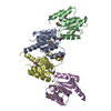
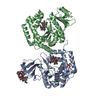
- PDB-2fmt: METHIONYL-TRNAFMET FORMYLTRANSFERASE COMPLEXED WITH FORMYL-METHIO... -
+ Open data

- Basic information
| Entry | Database: PDB / ID: 2fmt | ||||||
|---|---|---|---|---|---|---|---|
| Title | METHIONYL-TRNAFMET FORMYLTRANSFERASE COMPLEXED WITH FORMYL-METHIONYL-TRNAFMET | ||||||
 Components Components |
| ||||||
 Keywords Keywords | COMPLEX (METHYLTRANSFERASE/TRNA) / COMPLEX (METHYLTRANSFERASE-TRNA) /  FORMYLTRANSFERASE / INITIATION OF TRANSLATION / COMPLEX (METHYLTRANSFERASE-TRNA) complex FORMYLTRANSFERASE / INITIATION OF TRANSLATION / COMPLEX (METHYLTRANSFERASE-TRNA) complex | ||||||
| Function / homology |  Function and homology information Function and homology informationcharged-tRNA amino acid modification / methionyl-tRNA formyltransferase / conversion of methionyl-tRNA to N-formyl-methionyl-tRNA / methionyl-tRNA formyltransferase activity / cytosolSimilarity search - Function | ||||||
| Biological species |  Escherichia coli (E. coli) Escherichia coli (E. coli)synthetic construct (others) | ||||||
| Method | X-RAY DIFFRACTION / SYNCHROTRON / SIR / Resolution: 2.8 Å | ||||||
 Authors Authors | Schmitt, E. / Mechulam, Y. / Blanquet, S. | ||||||
 Citation Citation | Journal: EMBO J. / Year: 1998 Title: Crystal structure of methionyl-tRNAfMet transformylase complexed with the initiator formyl-methionyl-tRNAfMet. Authors: Schmitt, E. / Panvert, M. / Blanquet, S. / Mechulam, Y. | ||||||
| History |
|
- Structure visualization
| Structure viewer | Molecule: MolmilJmol/JSmol |
|---|
- Downloads & links
-Download
| PDBx/mmCIF format | 2fmt.cif.gz | 208.4 KB | Display | PDBx/mmCIF format |
|---|---|---|---|---|
| PDB format | pdb2fmt.ent.gz | 167.4 KB | Display | PDB format |
| PDBx/mmJSON format | 2fmt.json.gz | Tree view | PDBx/mmJSON format | |
| Others |  Other downloads Other downloads |
-Validation report
| Arichive directory | https://data.pdbj.org/pub/pdb/validation_reports/fm/2fmtftp://data.pdbj.org/pub/pdb/validation_reports/fm/2fmt | HTTPS FTP |
|---|
-Related structure data
| Related structure data | 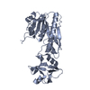 1fmtS S: Starting model for refinement |
|---|---|
| Similar structure data |


-Links
 PDBj
PDBj


- Assembly
| Deposited unit | 
| |||||||||||||||||||||
|---|---|---|---|---|---|---|---|---|---|---|---|---|---|---|---|---|---|---|---|---|---|---|
| 1 | 
| |||||||||||||||||||||
| 2 | 
| |||||||||||||||||||||
| Unit cell |
| |||||||||||||||||||||
| Noncrystallographic symmetry (NCS) | NCS domain:
NCS oper:
| |||||||||||||||||||||
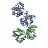
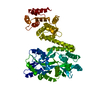
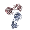
| Details | THE BIOLOGICALLY ACTIVE COMPLEX CORRESPONDS TO EITHER CHAINS A AND C, OR CHAINS B AND D. THE COORDINATES FOR CHAINS A AND C ARE MORE ACCURATE. |
-Components
| #1: RNA chain | Mass: 24832.918 Da / Num. of mol.: 2 / Source method: obtained synthetically / Source: (synth.) synthetic construct (others) #2: Protein | Mass: 34071.168 Da / Num. of mol.: 2 Source method: isolated from a genetically manipulated source Source: (gene. exp.) Escherichia coli (E. coli) / Strain: K37 / Cellular location: CYTOPLASM / Gene: FMT / Plasmid: PUCFATG / Cellular location (production host): CYTOPLASM / Production host: Escherichia coli (E. coli) / Strain (production host): JM101TRReferences: UniProt: P23882, methionyl-tRNA formyltransferase#3: Chemical |   Mass: 24.305 Da / Num. of mol.: 2 / Source method: obtained synthetically / Formula: Mg Mass: 24.305 Da / Num. of mol.: 2 / Source method: obtained synthetically / Formula: Mg#4: Chemical | N-Formylmethionine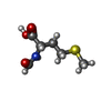  Type: L-peptide linking / Mass: 177.221 Da / Num. of mol.: 2 / Source method: obtained synthetically / Formula: C6H11NO3S Type: L-peptide linking / Mass: 177.221 Da / Num. of mol.: 2 / Source method: obtained synthetically / Formula: C6H11NO3S#5: Water | ChemComp-HOH / | Water Mass: 18.015 Da / Num. of mol.: 83 / Source method: isolated from a natural source / Formula: H2O Mass: 18.015 Da / Num. of mol.: 83 / Source method: isolated from a natural source / Formula: H2O |
|---|
-Experimental details
-Experiment
| Experiment | Method: X-RAY DIFFRACTION / Number of used crystals: 1 |
|---|
- Sample preparation
| Crystal | Density Matthews: 2.51 Å3/Da / Density % sol: 48 % | ||||||||||||||||||||||||||||||||||||||||||||||||||||||
|---|---|---|---|---|---|---|---|---|---|---|---|---|---|---|---|---|---|---|---|---|---|---|---|---|---|---|---|---|---|---|---|---|---|---|---|---|---|---|---|---|---|---|---|---|---|---|---|---|---|---|---|---|---|---|---|
| Crystal grow | pH: 6.6 / Details: pH 6.6 | ||||||||||||||||||||||||||||||||||||||||||||||||||||||
| Crystal | *PLUS | ||||||||||||||||||||||||||||||||||||||||||||||||||||||
| Crystal grow | *PLUS Method: unknown / Details: used to seeding | ||||||||||||||||||||||||||||||||||||||||||||||||||||||
| Components of the solutions | *PLUS
|
-Data collection
| Diffraction | Mean temperature: 100 K |
|---|---|
| Diffraction source | Source: SYNCHROTRON / Site: LURE  / Beamline: DW32 / Wavelength: 1 / Beamline: DW32 / Wavelength: 1 |
| Detector | Type: MARRESEARCH / Detector: IMAGE PLATE |
| Radiation | Monochromator: CRYSTAL / Monochromatic (M) / Laue (L): M / Scattering type: x-ray |
| Radiation wavelength | Wavelength: 1 Å / Relative weight: 1 |
| Reflection | Resolution: 2.8→30 Å / Num. obs: 29912 / % possible obs: 99.4 % / Redundancy: 3.3 % / Biso Wilson estimate: 78 Å2 / Rsym value: 0.051 / Net I/σ(I): 10.9 |
| Reflection shell | Resolution: 2.8→2.95 Å / Redundancy: 3.3 % / Mean I/σ(I) obs: 2.2 / Rsym value: 0.351 / % possible all: 99.6 |
| Reflection | *PLUS Rmerge(I) obs: 0.051 |
| Reflection shell | *PLUS Rmerge(I) obs: 0.35 |
- Processing
| Software |
| ||||||||||||||||||||||||||||||||||||||||||||||||||||||||||||||||||||||||||||||||
|---|---|---|---|---|---|---|---|---|---|---|---|---|---|---|---|---|---|---|---|---|---|---|---|---|---|---|---|---|---|---|---|---|---|---|---|---|---|---|---|---|---|---|---|---|---|---|---|---|---|---|---|---|---|---|---|---|---|---|---|---|---|---|---|---|---|---|---|---|---|---|---|---|---|---|---|---|---|---|---|---|---|
| Refinement | Method to determine structure: SIR Starting model: PDB ENTRY 1FMT Resolution: 2.8→19 Å / Isotropic thermal model: RESTRAINED / Cross valid method: THROUGHOUT / σ(F): 2 Details: THE POSITIONS OF BASES 1, 16 - 18 AND 37 IN CHAINS C AND D ARE TENTATIVE.
| ||||||||||||||||||||||||||||||||||||||||||||||||||||||||||||||||||||||||||||||||
| Solvent computation | Solvent model: FLAT MODEL / Bsol: 15.1 Å2 / ksol: 0.234 e/Å3 | ||||||||||||||||||||||||||||||||||||||||||||||||||||||||||||||||||||||||||||||||
| Displacement parameters | Biso mean: 62.7 Å2
| ||||||||||||||||||||||||||||||||||||||||||||||||||||||||||||||||||||||||||||||||
| Refinement step | Cycle: LAST / Resolution: 2.8→19 Å
| ||||||||||||||||||||||||||||||||||||||||||||||||||||||||||||||||||||||||||||||||
| Refine LS restraints |
| ||||||||||||||||||||||||||||||||||||||||||||||||||||||||||||||||||||||||||||||||
| Refine LS restraints NCS |
| ||||||||||||||||||||||||||||||||||||||||||||||||||||||||||||||||||||||||||||||||
| LS refinement shell | Resolution: 2.8→2.93 Å / Total num. of bins used: 8
| ||||||||||||||||||||||||||||||||||||||||||||||||||||||||||||||||||||||||||||||||
| Xplor file |
| ||||||||||||||||||||||||||||||||||||||||||||||||||||||||||||||||||||||||||||||||
| Software | *PLUS Name: CNS / Version: 0.3C / Classification: refinement | ||||||||||||||||||||||||||||||||||||||||||||||||||||||||||||||||||||||||||||||||
| Refine LS restraints | *PLUS
|
