 Movie
Movie Controller

[English] 日本語
 Yorodumi
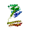
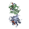
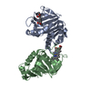
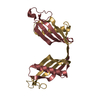
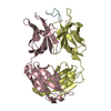
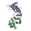
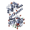
- PDB-1v8q: Crystal structure of ribosomal protein L27 from Thermus thermophi... -
+ Open data

- Basic information
| Entry | Database: PDB / ID: 1v8q | ||||||
|---|---|---|---|---|---|---|---|
| Title | Crystal structure of ribosomal protein L27 from Thermus thermophilus HB8 | ||||||
 Components Components | TT0826 | ||||||
 Keywords Keywords |  TRANSLATION / structural genomics / proteomics / RIKEN Structural Genomics/Proteomics Initiative / RSGI TRANSLATION / structural genomics / proteomics / RIKEN Structural Genomics/Proteomics Initiative / RSGI | ||||||
| Function / homology |  Function and homology informationribosome / structural constituent of ribosome / ribonucleoprotein complex / translation Function and homology informationribosome / structural constituent of ribosome / ribonucleoprotein complex / translationSimilarity search - Function | ||||||
| Biological species |   Thermus thermophilus (bacteria) Thermus thermophilus (bacteria) | ||||||
| Method | X-RAY DIFFRACTION / SYNCHROTRON / MAD / Resolution: 2.8 Å | ||||||
 Authors Authors | Wang, H. / Takemoto-Hori, C. / Murayama, K. / Terada, T. / Shirouzu, M. / Kuramitsu, S. / Yokoyama, S. / RIKEN Structural Genomics/Proteomics Initiative (RSGI) | ||||||
 Citation Citation | Journal: Protein Sci. / Year: 2004 Title: Crystal structure of ribosomal protein L27 from Thermus thermophilus HB8 Authors: Wang, H. / Takemoto-Hori, C. / Murayama, K. / Sakai, H. / Tatsuguchi, A. / Terada, T. / Shirouzu, M. / Kuramitsu, S. / Yokoyama, S. | ||||||
| History |
|
- Structure visualization
| Structure viewer | Molecule: MolmilJmol/JSmol |
|---|
- Downloads & links
-Download
| PDBx/mmCIF format | 1v8q.cif.gz | 63.9 KB | Display | PDBx/mmCIF format |
|---|---|---|---|---|
| PDB format | pdb1v8q.ent.gz | 47.8 KB | Display | PDB format |
| PDBx/mmJSON format | 1v8q.json.gz | Tree view | PDBx/mmJSON format | |
| Others |  Other downloads Other downloads |
-Validation report
| Arichive directory | https://data.pdbj.org/pub/pdb/validation_reports/v8/1v8qftp://data.pdbj.org/pub/pdb/validation_reports/v8/1v8q | HTTPS FTP |
|---|
-Related structure data
| Similar structure data | |
|---|---|
| Other databases |


-Links
 PDBj
PDBj


- Assembly
| Deposited unit | 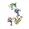
| ||||||||
|---|---|---|---|---|---|---|---|---|---|
| 1 |
| ||||||||
| Unit cell |
|
-Components
| #1: Protein | Mass: 9529.074 Da / Num. of mol.: 4 Source method: isolated from a genetically manipulated source Source: (gene. exp.) Thermus thermophilus (bacteria) / Plasmid: pET-26 / Production host: Escherichia coli (E. coli) / References: UniProt: P84123, UniProt: P60493*PLUS#2: Chemical | ChemComp-DTT / | Dithiothreitol  Mass: 154.251 Da / Num. of mol.: 1 / Source method: obtained synthetically / Formula: C4H10O2S2 Mass: 154.251 Da / Num. of mol.: 1 / Source method: obtained synthetically / Formula: C4H10O2S2#3: Water | ChemComp-HOH / | Water Mass: 18.015 Da / Num. of mol.: 18 / Source method: isolated from a natural source / Formula: H2O Mass: 18.015 Da / Num. of mol.: 18 / Source method: isolated from a natural source / Formula: H2O |
|---|
-Experimental details
-Experiment
| Experiment | Method: X-RAY DIFFRACTION / Number of used crystals: 2 |
|---|
- Sample preparation
| Crystal | Density Matthews: 2.15 Å3/Da / Density % sol: 42.78 % |
|---|---|
| Crystal grow | Temperature: 303 K / Method: vapor diffusion, hanging drop / pH: 6 Details: PEG 4000, Ammonium Acetate, tri-Sodium Citrate dihydrate, pH 6.0, VAPOR DIFFUSION, HANGING DROP, temperature 303K |
-Data collection
| Diffraction |
| ||||||||||||||||||
|---|---|---|---|---|---|---|---|---|---|---|---|---|---|---|---|---|---|---|---|
| Diffraction source |
| ||||||||||||||||||
| Detector |
| ||||||||||||||||||
| Radiation |
| ||||||||||||||||||
| Radiation wavelength |
| ||||||||||||||||||
| Reflection | Resolution: 2.8→45.83 Å / Num. obs: 7947 / % possible obs: 99.7 % / Observed criterion σ(F): -3 / Redundancy: 7.5 % / Biso Wilson estimate: 48.7 Å2 / Rsym value: 0.074 / Net I/σ(I): 27.8 | ||||||||||||||||||
| Reflection shell | Resolution: 2.8→2.9 Å / Redundancy: 6.4 % / Mean I/σ(I) obs: 3.6 / Num. unique all: 766 / Rsym value: 0.43 / % possible all: 97.3 |

- Processing
| Software |
| ||||||||||||||||||||
|---|---|---|---|---|---|---|---|---|---|---|---|---|---|---|---|---|---|---|---|---|---|
| Refinement | Method to determine structure: MAD / Resolution: 2.8→45.83 Å / Rfactor Rfree error: 0.008 / Data cutoff high absF: 237937.01 / Data cutoff low absF: 0 / Isotropic thermal model: RESTRAINED / Cross valid method: THROUGHOUT / σ(F): 0
| ||||||||||||||||||||
| Solvent computation | Solvent model: FLAT MODEL / Bsol: 58.2951 Å2 / ksol: 0.389673 e/Å3 | ||||||||||||||||||||
| Displacement parameters | Biso mean: 58.9 Å2
| ||||||||||||||||||||
| Refine analyze |
| ||||||||||||||||||||
| Refinement step | Cycle: LAST / Resolution: 2.8→45.83 Å
| ||||||||||||||||||||
| Refine LS restraints |
| ||||||||||||||||||||
| LS refinement shell | Resolution: 2.8→2.98 Å / Rfactor Rfree error: 0.035 / Total num. of bins used: 6
| ||||||||||||||||||||
| Xplor file |
|
