 Movie
Movie Controller


+ Open data

- Basic information
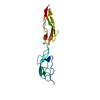
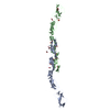
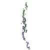
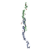
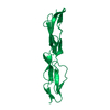
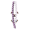
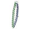
| Entry | Database: PDB / ID: 1uot | ||||||
|---|---|---|---|---|---|---|---|
| Title | HUMAN CD55 DOMAINS 3 & 4 | ||||||
 Components Components | COMPLEMENT DECAY-ACCELERATING FACTOR | ||||||
 Keywords Keywords | REGULATOR OF COMPLEMENT PATHWAY / IMMUNE SYSTEM PROTEIN / COMPLEMENT DECAY ACCELERATING FACTOR / ENTEROVIRAL RECEPTOR / BACTERIAL RECEPTOR / LIGAND FOR CD97 / COMPLEMENT PATHWAY / ALTERNATIVE SPLICING / GPI-ANCHOR | ||||||
| Function / homology |  Function and homology information Function and homology informationregulation of lipopolysaccharide-mediated signaling pathway / negative regulation of complement activation / regulation of complement-dependent cytotoxicity / regulation of complement activation / respiratory burst / positive regulation of CD4-positive, alpha-beta T cell activation / positive regulation of CD4-positive, alpha-beta T cell proliferation / Class B/2 (Secretin family receptors) / ficolin-1-rich granule membrane / side of membrane ...regulation of lipopolysaccharide-mediated signaling pathway / negative regulation of complement activation / regulation of complement-dependent cytotoxicity / regulation of complement activation / respiratory burst / positive regulation of CD4-positive, alpha-beta T cell activation / positive regulation of CD4-positive, alpha-beta T cell proliferation / Class B/2 (Secretin family receptors) / ficolin-1-rich granule membrane / side of membrane / COPI-mediated anterograde transport / transport vesicle / endoplasmic reticulum-Golgi intermediate compartment membrane / complement activation, classical pathway / secretory granule membrane / Regulation of Complement cascade / positive regulation of T cell cytokine production / virus receptor activity / positive regulation of cytosolic calcium ion concentration / membrane raft / Golgi membrane / innate immune response / lipid binding / Neutrophil degranulation / cell surface / extracellular exosome / extracellular region / plasma membrane Similarity search - Function | ||||||
| Biological species |  HOMO SAPIENS (human) HOMO SAPIENS (human) | ||||||
| Method |  X-RAY DIFFRACTION / SYNCHROTRON / MOLECULAR REPLACEMENT / Resolution: 3 Å X-RAY DIFFRACTION / SYNCHROTRON / MOLECULAR REPLACEMENT / Resolution: 3 Å | ||||||
 Authors Authors | Williams, P. / Chaudhry, Y. / Goodfellow, I.G. / Billington, J. / Spiller, B. / Evans, D.J. / Lea, S.M. | ||||||
 Citation Citation | Journal: J.Biol.Chem. / Year: 2003 Title: Mapping Cd55 Function. The Structure of Two Pathogen-Binding Domains at 1.7 A Authors: Williams, P. / Chaudhry, Y. / Goodfellow, I.G. / Billington, J. / Powell, R. / Spiller, O.B. / Evans, D.J. / Lea, S.M. #1: Journal: Acta Crystallogr.,Sect.D / Year: 1999 Title: Crystallization and Preliminary X-Ray Diffraction Analysis of a Biologically Active Fragment of Cd55 Authors: Lea, S.M. / Powell, R. / Evans, D.J. #2: Journal: J.Biol.Chem. / Year: 1998 Title: Determination of the Affinity and Kinetic Constants for the Interaction between the Human Virus Echovirus 11 and its Cellular Receptor, Cd55 Authors: Lea, S.M. / Powell, R. / Mckee, T. / Evans, D.J. / Brown, D.J. / Stuart, D.I. / Van Der Merwe, A. | ||||||
| History |
|
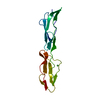
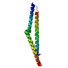
- Structure visualization
| Structure viewer | Molecule: MolmilJmol/JSmol |
|---|
- Downloads & links
-Download
| PDBx/mmCIF format | 1uot.cif.gz | 36.6 KB | Display | PDBx/mmCIF format |
|---|---|---|---|---|
| PDB format | pdb1uot.ent.gz | 24 KB | Display | PDB format |
| PDBx/mmJSON format | 1uot.json.gz | Tree view | PDBx/mmJSON format | |
| Others |  Other downloads Other downloads |
-Validation report
| Summary document | 1uot_validation.pdf.gz | 428 KB | Display | wwPDB validaton report |
|---|---|---|---|---|
| Full document | 1uot_full_validation.pdf.gz | 431.8 KB | Display | |
| Data in XML | 1uot_validation.xml.gz | 7.2 KB | Display | |
| Data in CIF | 1uot_validation.cif.gz | 8.5 KB | Display | |
| Arichive directory | https://data.pdbj.org/pub/pdb/validation_reports/uo/1uotftp://data.pdbj.org/pub/pdb/validation_reports/uo/1uot | HTTPS FTP |
-Related structure data
| Related structure data |  1h03SC  1h04C 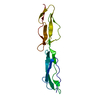 1h2pC 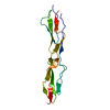 1h2qC S: Starting model for refinement C: citing same article ( |
|---|---|
| Similar structure data |


-Links
 PDBj
PDBj


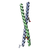
- Assembly
| Deposited unit | 
| ||||||||
|---|---|---|---|---|---|---|---|---|---|
| 1 |
| ||||||||
| Unit cell |
|
-Components
| #1: Protein | Mass: 13586.049 Da / Num. of mol.: 1 / Fragment: EXTRACELLULAR SCR DOMAINS 3 & 4, RESIDUES 161-285 Source method: isolated from a genetically manipulated source Source: (gene. exp.) HOMO SAPIENS (human) / Production host:  PICHIA PASTORIS (fungus) / References: UniProt: P08174 PICHIA PASTORIS (fungus) / References: UniProt: P08174 |
|---|---|
| #2: Water | ChemComp-HOH /  Mass: 18.015 Da / Num. of mol.: 8 / Source method: isolated from a natural source / Formula: H2O Mass: 18.015 Da / Num. of mol.: 8 / Source method: isolated from a natural source / Formula: H2O |
| Compound details | FUNCTION: THIS PROTEIN RECOGNIZES C4B AND C3B FRAGMENTS THAT CONDENSE WITH CELL-SURFACE HYDROXYL OR ...FUNCTION: THIS PROTEIN RECOGNIZES |
-Experimental details
-Experiment
| Experiment | Method: X-RAY DIFFRACTION / Number of used crystals: 1 |
|---|
- Sample preparation
| Crystal | Density Matthews: 2.28 Å3/Da / Density % sol: 45 % |
|---|---|
| Crystal grow | pH: 5.6 / Details: pH 5.60 |
-Data collection
| Diffraction | Mean temperature: 100 K |
|---|---|
| Diffraction source | Source: SYNCHROTRON / Site: SRS  / Beamline: PX9.6 / Wavelength: 0.87 / Beamline: PX9.6 / Wavelength: 0.87 |
| Detector | Type: MARRESEARCH / Detector: IMAGE PLATE |
| Radiation | Protocol: SINGLE WAVELENGTH / Monochromatic (M) / Laue (L): M / Scattering type: x-ray |
| Radiation wavelength | Wavelength: 0.87 Å / Relative weight: 1 |
| Reflection | Resolution: 3→40 Å / Num. obs: 2432 / % possible obs: 88 % / Redundancy: 4.8 % / Biso Wilson estimate: 4.123 Å2 / Rmerge(I) obs: 0.12 / Net I/σ(I): 2.3 |
| Reflection shell | Resolution: 3→3.1 Å / Rmerge(I) obs: 0.28 / Mean I/σ(I) obs: 1 / % possible all: 82 |
- Processing
| Software |
| |||||||||||||||||||||||||||||||||||||||||||||||||||||||||||||||||||||||||||||||||||||||||||||||
|---|---|---|---|---|---|---|---|---|---|---|---|---|---|---|---|---|---|---|---|---|---|---|---|---|---|---|---|---|---|---|---|---|---|---|---|---|---|---|---|---|---|---|---|---|---|---|---|---|---|---|---|---|---|---|---|---|---|---|---|---|---|---|---|---|---|---|---|---|---|---|---|---|---|---|---|---|---|---|---|---|---|---|---|---|---|---|---|---|---|---|---|---|---|---|---|---|
| Refinement | Method to determine structure: MOLECULAR REPLACEMENT Starting model: PDB ENTRY 1H03 Resolution: 3→18.5 Å / Isotropic thermal model: TNT BCORREL / Cross valid method: THROUGHOUT / σ(F): 0 / Stereochemistry target values: TNT PROTGEO Details: DRIVEN BY BUSTER MAXIMUM LIKELIHOOD. THIS IS A NEW REFINEMENT OF PDB ENTRY 1H2Q
| |||||||||||||||||||||||||||||||||||||||||||||||||||||||||||||||||||||||||||||||||||||||||||||||
| Solvent computation | Solvent model: BABINET SCALING / Bsol: 28 Å2 / ksol: 1.14 e/Å3 | |||||||||||||||||||||||||||||||||||||||||||||||||||||||||||||||||||||||||||||||||||||||||||||||
| Refinement step | Cycle: LAST / Resolution: 3→18.5 Å
| |||||||||||||||||||||||||||||||||||||||||||||||||||||||||||||||||||||||||||||||||||||||||||||||
| Refine LS restraints |
|
