 Movie
Movie Controller


+ Open data

- Basic information
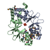
| Entry | Database: PDB / ID: 1s7z | ||||||
|---|---|---|---|---|---|---|---|
| Title | Structure of Ocr from Bacteriophage T7 | ||||||
 Components Components | Gene 0.3 protein | ||||||
 Keywords Keywords |  GENE REGULATION / all helical GENE REGULATION / all helical | ||||||
| Function / homology |  Function and homology information Function and homology information | ||||||
| Biological species |   Enterobacteria phage T7 (virus) Enterobacteria phage T7 (virus) | ||||||
| Method | X-RAY DIFFRACTION / SYNCHROTRON / MAD / Resolution: 1.83 Å | ||||||
 Authors Authors | Walkinshaw, M.D. / Taylor, P. / Sturrock, S.S. / Atanasiu, C. / Berg, T. / Henderson, R.M. / Edwardson, J.M. / Dryden, D.T. | ||||||
 Citation Citation | Journal: Mol.Cell / Year: 2002 Title: Structure of Ocr from Bacteriophage T7, a Protein that Mimics B-Form DNA Authors: Walkinshaw, M.D. / Taylor, P. / Sturrock, S.S. / Atanasiu, C. / Berg, T. / Henderson, R.M. / Edwardson, J.M. / Dryden, D.T. | ||||||
| History |
|
- Structure visualization
| Structure viewer | Molecule: MolmilJmol/JSmol |
|---|
- Downloads & links
-Download
| PDBx/mmCIF format | 1s7z.cif.gz | 35.1 KB | Display | PDBx/mmCIF format |
|---|---|---|---|---|
| PDB format | pdb1s7z.ent.gz | 26.9 KB | Display | PDB format |
| PDBx/mmJSON format | 1s7z.json.gz | Tree view | PDBx/mmJSON format | |
| Others |  Other downloads Other downloads |
-Validation report
| Arichive directory | https://data.pdbj.org/pub/pdb/validation_reports/s7/1s7zftp://data.pdbj.org/pub/pdb/validation_reports/s7/1s7z | HTTPS FTP |
|---|
-Related structure data
| Similar structure data |
|---|


-Links
 PDBj
PDBj- Assembly
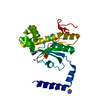
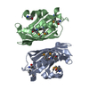
| Deposited unit | 
| ||||||||
|---|---|---|---|---|---|---|---|---|---|
| 1 | 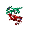
| ||||||||
| 2 | 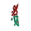
| ||||||||
| 3 |
| ||||||||
| Unit cell |
| ||||||||
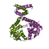
| Details | The tight (sub nanomolar) dimer in solution is retained in the crystal by a crystallographic two-fold axis The interface is only 250 Angstroms squared for each monomer |
-Components
| #1: Protein | Mass: 14100.386 Da / Num. of mol.: 1 Source method: isolated from a genetically manipulated source Source: (gene. exp.) Enterobacteria phage T7 (virus) / Genus: T7-like viruses / Gene: 0.3 / Production host:  Escherichia coli (E. coli) / References: UniProt: P03775 Escherichia coli (E. coli) / References: UniProt: P03775 | ||
|---|---|---|---|
| #2: Chemical |   Mass: 132.905 Da / Num. of mol.: 3 / Source method: obtained synthetically / Formula: Cs Mass: 132.905 Da / Num. of mol.: 3 / Source method: obtained synthetically / Formula: Cs#3: Water | ChemComp-HOH / | Water Mass: 18.015 Da / Num. of mol.: 118 / Source method: isolated from a natural source / Formula: H2O Mass: 18.015 Da / Num. of mol.: 118 / Source method: isolated from a natural source / Formula: H2O |
-Experimental details
-Experiment
| Experiment | Method: X-RAY DIFFRACTION / Number of used crystals: 1 |
|---|
- Sample preparation
| Crystal | Density Matthews: 1.85 Å3/Da / Density % sol: 33.39 % | ||||||||||||||||||||||||||||||||||||||||||
|---|---|---|---|---|---|---|---|---|---|---|---|---|---|---|---|---|---|---|---|---|---|---|---|---|---|---|---|---|---|---|---|---|---|---|---|---|---|---|---|---|---|---|---|
| Crystal grow | Temperature: 291 K / Method: vapor diffusion, hanging drop / pH: 8.2 Details: 32%PEG-8000, 0.1M Tris-HCl, 0.2M caesium chloride, pH 8.2, VAPOR DIFFUSION, HANGING DROP, temperature 291K | ||||||||||||||||||||||||||||||||||||||||||
| Crystal grow | *PLUS Temperature: 291 K / Method: vapor diffusion, hanging drop / Details: Sturrock, S.S., (2001) Acta Cryst., D57, 1652. | ||||||||||||||||||||||||||||||||||||||||||
| Components of the solutions | *PLUS
|
-Data collection
| Diffraction | Mean temperature: 100 K | |||||||||||||||
|---|---|---|---|---|---|---|---|---|---|---|---|---|---|---|---|---|
| Diffraction source | Source: SYNCHROTRON / Site: SRS  / Beamline: PX14.2 / Wavelength: 0.87 / Wavelength: 1.2, 0.9777,0.9793 / Beamline: PX14.2 / Wavelength: 0.87 / Wavelength: 1.2, 0.9777,0.9793 | |||||||||||||||
| Detector | Type: MARRESEARCH / Detector: CCD / Date: Mar 1, 2000 / Details: channel cut monochromator | |||||||||||||||
| Radiation | Monochromator: Silicon / Protocol: MAD / Monochromatic (M) / Laue (L): M / Scattering type: x-ray | |||||||||||||||
| Radiation wavelength |
| |||||||||||||||
| Reflection | Resolution: 1.83→38.9 Å / Num. all: 8923 / Num. obs: 8923 / Observed criterion σ(F): 0 / Observed criterion σ(I): 0 / Redundancy: 8 % / Rmerge(I) obs: 0.049 / Rsym value: 0.049 / Net I/σ(I): 17.9 | |||||||||||||||
| Reflection shell | Resolution: 1.83→1.88 Å / Rmerge(I) obs: 0.096 / Mean I/σ(I) obs: 13.3 / Rsym value: 0.096 / % possible all: 98.5 | |||||||||||||||
| Reflection | *PLUS Highest resolution: 1.8 Å / % possible obs: 99.3 % / Num. measured all: 71916 | |||||||||||||||
| Reflection shell | *PLUS % possible obs: 98.5 % |
- Processing
| Software |
| ||||||||||||||||||||||||||||||||||||||||||||||||||||||||||||||||||||||||||||||||||||||||||||||||||||||||||||||||||||||||||||||||||||||||||||||||||||||||||||||||
|---|---|---|---|---|---|---|---|---|---|---|---|---|---|---|---|---|---|---|---|---|---|---|---|---|---|---|---|---|---|---|---|---|---|---|---|---|---|---|---|---|---|---|---|---|---|---|---|---|---|---|---|---|---|---|---|---|---|---|---|---|---|---|---|---|---|---|---|---|---|---|---|---|---|---|---|---|---|---|---|---|---|---|---|---|---|---|---|---|---|---|---|---|---|---|---|---|---|---|---|---|---|---|---|---|---|---|---|---|---|---|---|---|---|---|---|---|---|---|---|---|---|---|---|---|---|---|---|---|---|---|---|---|---|---|---|---|---|---|---|---|---|---|---|---|---|---|---|---|---|---|---|---|---|---|---|---|---|---|---|---|---|
| Refinement | Method to determine structure: MAD / Resolution: 1.83→38.9 Å / Cor.coef. Fo:Fc: 0.94 / Cor.coef. Fo:Fc free: 0.888 / SU B: 4.209 / SU ML: 0.132 / Cross valid method: THROUGHOUT / ESU R: 0.152 / ESU R Free: 0.155 / Stereochemistry target values: MAXIMUM LIKELIHOOD / Details: HYDROGENS HAVE BEEN ADDED IN THE RIDING POSITIONS
| ||||||||||||||||||||||||||||||||||||||||||||||||||||||||||||||||||||||||||||||||||||||||||||||||||||||||||||||||||||||||||||||||||||||||||||||||||||||||||||||||
| Solvent computation | Ion probe radii: 0.8 Å / Shrinkage radii: 0.8 Å / VDW probe radii: 1.4 Å / Solvent model: BABINET MODEL WITH MASK | ||||||||||||||||||||||||||||||||||||||||||||||||||||||||||||||||||||||||||||||||||||||||||||||||||||||||||||||||||||||||||||||||||||||||||||||||||||||||||||||||
| Displacement parameters | Biso mean: 10.834 Å2
| ||||||||||||||||||||||||||||||||||||||||||||||||||||||||||||||||||||||||||||||||||||||||||||||||||||||||||||||||||||||||||||||||||||||||||||||||||||||||||||||||
| Refinement step | Cycle: LAST / Resolution: 1.83→38.9 Å
| ||||||||||||||||||||||||||||||||||||||||||||||||||||||||||||||||||||||||||||||||||||||||||||||||||||||||||||||||||||||||||||||||||||||||||||||||||||||||||||||||
| Refine LS restraints |
| ||||||||||||||||||||||||||||||||||||||||||||||||||||||||||||||||||||||||||||||||||||||||||||||||||||||||||||||||||||||||||||||||||||||||||||||||||||||||||||||||
| LS refinement shell | Resolution: 1.83→1.879 Å / Total num. of bins used: 20 /
| ||||||||||||||||||||||||||||||||||||||||||||||||||||||||||||||||||||||||||||||||||||||||||||||||||||||||||||||||||||||||||||||||||||||||||||||||||||||||||||||||
| Software | *PLUS Version: 5 / Classification: refinement | ||||||||||||||||||||||||||||||||||||||||||||||||||||||||||||||||||||||||||||||||||||||||||||||||||||||||||||||||||||||||||||||||||||||||||||||||||||||||||||||||
| Refinement | *PLUS Rfactor Rfree: 0.241 / Rfactor Rwork: 0.174 | ||||||||||||||||||||||||||||||||||||||||||||||||||||||||||||||||||||||||||||||||||||||||||||||||||||||||||||||||||||||||||||||||||||||||||||||||||||||||||||||||
| Solvent computation | *PLUS | ||||||||||||||||||||||||||||||||||||||||||||||||||||||||||||||||||||||||||||||||||||||||||||||||||||||||||||||||||||||||||||||||||||||||||||||||||||||||||||||||
| Displacement parameters | *PLUS | ||||||||||||||||||||||||||||||||||||||||||||||||||||||||||||||||||||||||||||||||||||||||||||||||||||||||||||||||||||||||||||||||||||||||||||||||||||||||||||||||
| Refine LS restraints | *PLUS
|
