 Movie
Movie Controller

[English] 日本語
 Yorodumi
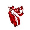
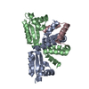
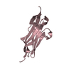
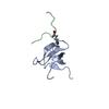
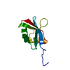
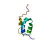
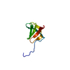
- PDB-1rqs: NMR structure of C-terminal domain of ribosomal protein L7 from E.coli -
+ Open data

- Basic information
| Entry | Database: PDB / ID: 1rqs | ||||||
|---|---|---|---|---|---|---|---|
| Title | NMR structure of C-terminal domain of ribosomal protein L7 from E.coli | ||||||
 Components Components | 50S ribosomal protein L7/L12 Ribosome Ribosome | ||||||
 Keywords Keywords | RIBOSOME / protein L7/L12 | ||||||
| Function / homology |  Function and homology informationribosome binding / large ribosomal subunit / cytoplasmic translation / cytosolic large ribosomal subunit / structural constituent of ribosome / translation / mRNA binding / protein homodimerization activity / cytosol / cytoplasm Function and homology informationribosome binding / large ribosomal subunit / cytoplasmic translation / cytosolic large ribosomal subunit / structural constituent of ribosome / translation / mRNA binding / protein homodimerization activity / cytosol / cytoplasmSimilarity search - Function | ||||||
| Biological species |  Escherichia coli (E. coli)Escherichia coli O6 (bacteria)Escherichia coli O157:H7 (bacteria)Shigella flexneri (bacteria) Escherichia coli (E. coli)Escherichia coli O6 (bacteria)Escherichia coli O157:H7 (bacteria)Shigella flexneri (bacteria) | ||||||
| Method | SOLUTION NMR / simulated annealing, molecular dynamics in torsion angle space | ||||||
 Authors Authors | Bocharov, E.V. / Sobol, A.G. / Pavlov, K.V. / Korzhnev, D.M. / Jaravine, V.A. / Gudkov, A.T. / Arseniev, A.S. | ||||||
 Citation Citation | Journal: J.Biol.Chem. / Year: 2004 Title: From structure and dynamics of protein L7/L12 to molecular switching in ribosome. Authors: Bocharov, E.V. / Sobol, A.G. / Pavlov, K.V. / Korzhnev, D.M. / Jaravine, V.A. / Gudkov, A.T. / Arseniev, A.S. | ||||||
| History |
|
- Structure visualization
| Structure viewer | Molecule: MolmilJmol/JSmol |
|---|
- Downloads & links
-Download
| PDBx/mmCIF format | 1rqs.cif.gz | 417.1 KB | Display | PDBx/mmCIF format |
|---|---|---|---|---|
| PDB format | pdb1rqs.ent.gz | 363.8 KB | Display | PDB format |
| PDBx/mmJSON format | 1rqs.json.gz | Tree view | PDBx/mmJSON format | |
| Others |  Other downloads Other downloads |
-Validation report
| Arichive directory | https://data.pdbj.org/pub/pdb/validation_reports/rq/1rqsftp://data.pdbj.org/pub/pdb/validation_reports/rq/1rqs | HTTPS FTP |
|---|
-Related structure data
| Related structure data | 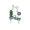 1rqtC 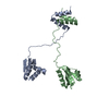 1rquC 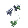 1rqvC C: citing same article ( |
|---|---|
| Similar structure data | |
| Other databases |


-Links
 PDBj
PDBj


- Assembly
| Deposited unit | 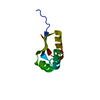
| |||||||||
|---|---|---|---|---|---|---|---|---|---|---|
| 1 |
| |||||||||
| NMR ensembles |
|
-Components
| #1: Protein | Ribosome / L8 Mass: 7573.641 Da / Num. of mol.: 1 / Fragment: C-Terminal Domain Source method: isolated from a genetically manipulated source Source: (gene. exp.) Escherichia coli, Escherichia coli O6, Escherichia coli O157:H7, Shigella flexneri Genus: Escherichia, Escherichia, Escherichia, Shigella / Species: , Escherichia coli, Escherichia coli, / Strain: , O6, O157:H7, / Plasmid: pPR1 / Production host: Escherichia coli (E. coli) / Strain (production host): XL1 / References: UniProt: P0A7K2 |
|---|
-Experimental details
-Experiment
| Experiment | Method: SOLUTION NMR | ||||||||||||||||||||||||
|---|---|---|---|---|---|---|---|---|---|---|---|---|---|---|---|---|---|---|---|---|---|---|---|---|---|
| NMR experiment |
| ||||||||||||||||||||||||
| NMR details | Text: The structure was determined using triple-resonance NMR spectroscopy |
- Sample preparation
| Details |
| ||||||||||||
|---|---|---|---|---|---|---|---|---|---|---|---|---|---|
| Sample conditions | Ionic strength: 0.15 / pH: 6.9 / Pressure: ambient / Temperature: 303 K | ||||||||||||
| Crystal grow | *PLUS Method: other / Details: NMR |
-NMR measurement
| Radiation | Protocol: SINGLE WAVELENGTH / Monochromatic (M) / Laue (L): M |
|---|---|
| Radiation wavelength | Relative weight: 1 |
| NMR spectrometer | Type: Varian UNITY / Manufacturer: Varian / Model: UNITY / Field strength: 600 MHz |
- Processing
| NMR software |
| ||||||||||||||||||||
|---|---|---|---|---|---|---|---|---|---|---|---|---|---|---|---|---|---|---|---|---|---|
| Refinement | Method: simulated annealing, molecular dynamics in torsion angle space Software ordinal: 1 | ||||||||||||||||||||
| NMR representative | Selection criteria: lowest energy | ||||||||||||||||||||
| NMR ensemble | Conformer selection criteria: structures with the least restraint violations,target function Conformers calculated total number: 200 / Conformers submitted total number: 20 |
