 Movie
Movie Controller


+ Open data

- Basic information
| Entry | Database: PDB / ID: 1pfo | ||||||
|---|---|---|---|---|---|---|---|
| Title | PERFRINGOLYSIN O | ||||||
 Components Components | PERFRINGOLYSIN O | ||||||
 Keywords Keywords |  TOXIN / THIOL-ACTIVATED CYTOLYSIN / HEMOLYSIS / CYTOLYSIS TOXIN / THIOL-ACTIVATED CYTOLYSIN / HEMOLYSIS / CYTOLYSIS | ||||||
| Function / homology |  Function and homology informationhemolysis in another organism / cholesterol binding / toxin activity / membrane => GO:0016020 / host cell plasma membrane / extracellular region / membrane Function and homology informationhemolysis in another organism / cholesterol binding / toxin activity / membrane => GO:0016020 / host cell plasma membrane / extracellular region / membraneSimilarity search - Function | ||||||
| Biological species |   Clostridium perfringens (bacteria) Clostridium perfringens (bacteria) | ||||||
| Method | X-RAY DIFFRACTION / SYNCHROTRON / MIR / Resolution: 2.2 Å | ||||||
 Authors Authors | Rossjohn, J. / Parker, M.W. | ||||||
 Citation Citation | Journal: Cell / Year: 1997 Title: Structure of a cholesterol-binding, thiol-activated cytolysin and a model of its membrane form. Authors: J Rossjohn / S C Feil / W J McKinstry / R K Tweten / M W Parker /  Abstract: The mechanisms by which proteins gain entry into membranes is a fundamental problem in biology. Here, we present the first crystal structure of a thiol-activated cytolysin, perfringolysin O, a member ...The mechanisms by which proteins gain entry into membranes is a fundamental problem in biology. Here, we present the first crystal structure of a thiol-activated cytolysin, perfringolysin O, a member of a large family of toxins that kill eukaryotic cells by punching holes in their membranes. The molecule adopts an unusually elongated shape rich in beta sheet. We have used electron microscopy data to construct a detailed model of the membrane channel form of the toxin. The structures reveal a novel mechanism for membrane insertion. Surprisingly, the toxin receptor, cholesterol, appears to play multiple roles: targeting, promotion of oligomerization, triggering a membrane insertion competent form, and stabilizing the membrane pore. | ||||||
| History |
|
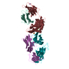
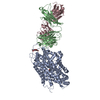
- Structure visualization
| Structure viewer | Molecule: MolmilJmol/JSmol |
|---|
- Downloads & links
-Download
| PDBx/mmCIF format | 1pfo.cif.gz | 112.1 KB | Display | PDBx/mmCIF format |
|---|---|---|---|---|
| PDB format | pdb1pfo.ent.gz | 86.1 KB | Display | PDB format |
| PDBx/mmJSON format | 1pfo.json.gz | Tree view | PDBx/mmJSON format | |
| Others |  Other downloads Other downloads |
-Validation report
| Arichive directory | https://data.pdbj.org/pub/pdb/validation_reports/pf/1pfoftp://data.pdbj.org/pub/pdb/validation_reports/pf/1pfo | HTTPS FTP |
|---|
-Related structure data
| Similar structure data |
|---|


-Links
 PDBj
PDBj- Assembly
| Deposited unit | 
| ||||||||
|---|---|---|---|---|---|---|---|---|---|
| 1 | 
| ||||||||
| Unit cell |
|
-Components
| #1: Protein | Mass: 55831.598 Da / Num. of mol.: 1 Source method: isolated from a genetically manipulated source Source: (gene. exp.) Clostridium perfringens (bacteria)Description: SECRETED AS A MONOMER, FORMS MEMBRANE-BOUND OLIGOMERS Cellular location: CYTOPLASM / Production host: Escherichia coli (E. coli) / References: UniProt: P19995, UniProt: P0C2E9*PLUS |
|---|---|
| #2: Water | ChemComp-HOH / Water Mass: 18.015 Da / Num. of mol.: 330 / Source method: isolated from a natural source / Formula: H2O Mass: 18.015 Da / Num. of mol.: 330 / Source method: isolated from a natural source / Formula: H2O |
-Experimental details
-Experiment
| Experiment | Method: X-RAY DIFFRACTION / Number of used crystals: 1 |
|---|
- Sample preparation
| Crystal | Density Matthews: 3.27 Å3/Da / Density % sol: 66 % | |||||||||||||||||||||||||
|---|---|---|---|---|---|---|---|---|---|---|---|---|---|---|---|---|---|---|---|---|---|---|---|---|---|---|
| Crystal grow | pH: 8.4 / Details: 10% PEG 20000, 100MM BICINE, PH 8.4, 2% DIOXANE | |||||||||||||||||||||||||
| Crystal grow | *PLUS Temperature: 22 ℃ / pH: 8.7 / Method: vapor diffusion, hanging drop / Details: Feil, S.C., (1996) FEBS Lett., 397, 290. | |||||||||||||||||||||||||
| Components of the solutions | *PLUS
|
-Data collection
| Diffraction | Mean temperature: 100 K |
|---|---|
| Diffraction source | Source: SYNCHROTRON / Site: Photon Factory  / Beamline: BL-6A / Wavelength: 1 / Beamline: BL-6A / Wavelength: 1 |
| Detector | Detector: FILM / Date: Apr 1, 1996 |
| Radiation | Monochromatic (M) / Laue (L): M / Scattering type: x-ray |
| Radiation wavelength | Wavelength: 1 Å / Relative weight: 1 |
| Reflection | Resolution: 2.1→20 Å / Num. obs: 37231 / % possible obs: 86 % / Redundancy: 3.26 % / Rmerge(I) obs: 0.058 / Net I/σ(I): 19.7 |
| Reflection shell | Resolution: 2.1→2.17 Å / Rmerge(I) obs: 0.41 / Mean I/σ(I) obs: 3.1 / % possible all: 87.1 |
- Processing
| Software |
| ||||||||||||||||||||||||||||||||||||||||||||||||||||||||||||
|---|---|---|---|---|---|---|---|---|---|---|---|---|---|---|---|---|---|---|---|---|---|---|---|---|---|---|---|---|---|---|---|---|---|---|---|---|---|---|---|---|---|---|---|---|---|---|---|---|---|---|---|---|---|---|---|---|---|---|---|---|---|
| Refinement | Method to determine structure: MIR / Resolution: 2.2→20 Å / Data cutoff high absF: 10000000 / Data cutoff low absF: 0.001 / Cross valid method: THROUGHOUT / σ(F): 0 Details: THE FIRST NINE RESIDUES HAVE HIGH MOBILITY, AND THE CORRESPONDING DENSITY WAS SMEARED OUT. THE R FREE WAS LOWER WHEN THESE RESIDUES WERE INCLUDED IN THE MODEL. UNCERTAINTY IN PLACEMENT AROUND RESIDUE 60.
| ||||||||||||||||||||||||||||||||||||||||||||||||||||||||||||
| Refinement step | Cycle: LAST / Resolution: 2.2→20 Å
| ||||||||||||||||||||||||||||||||||||||||||||||||||||||||||||
| Refine LS restraints |
| ||||||||||||||||||||||||||||||||||||||||||||||||||||||||||||
| LS refinement shell | Resolution: 2.2→2.3 Å / Total num. of bins used: 8
| ||||||||||||||||||||||||||||||||||||||||||||||||||||||||||||
| Software | *PLUS Name: X-PLOR / Version: 3.8 / Classification: refinement | ||||||||||||||||||||||||||||||||||||||||||||||||||||||||||||
| Refine LS restraints | *PLUS
|
